• Zirconia Pumps vs. Traditional Pump Materials A Comparative Guide

    |

    When selecting the right pump for industrial applications, choosing the correct material is essential to ensure durability, reliability, and cost-effectiveness. Pumps are used in various industries, from chemical processing and water treatment to the pharmaceutical and food sectors. While traditional pump materials such as stainless steel, plastic, and graphite have been the go-to options for many years, advanced materials like zirconia are gaining popularity for their superior performance, particularly in demanding environments. In this comparative guide, we will explore zirconia pumps and how they stack up against traditional pump materials in terms of durability, performance, and cost-effectiveness.

    What Are Zirconia Pumps?

    Zirconia pumps are made from zirconium oxide (ZrO2), a highly durable ceramic material known for its exceptional strength, hardness, and resistance to heat and corrosion. Zirconia is used in applications where traditional pump materials like stainless steel or plastic would fail due to high wear, chemical exposure, or extreme temperature conditions. These pumps are particularly valued in industries like chemical processing, water treatment, food production, and pharmaceuticals.

    Key Properties of Zirconia Pumps

    Before we compare zirconia pumps with traditional materials, it’s essential to understand the unique properties of zirconia that make it an attractive option for many industrial applications:

    1. Thermal Resistance: Zirconia can withstand extremely high temperatures, making it ideal for high-heat environments. It maintains structural integrity even at temperatures exceeding 1,000°C (1,832°F).

    2. Chemical Inertness: Zirconia is highly resistant to most acids, alkalis, and solvents, which makes it an excellent choice for industries where the pump comes into contact with aggressive chemicals.

    3. Wear Resistance: Zirconia’s hardness and toughness make it highly resistant to abrasion, erosion, and wear, which is essential in industries dealing with abrasive slurries or materials.

    4. Mechanical Strength: Zirconia possesses excellent mechanical strength, which means it can handle high pressure and physical stress without cracking or deforming.

    Comparison of Zirconia Pumps with Traditional Pump Materials

    Now that we understand the key properties of zirconia, let’s compare it with some of the most common traditional pump materials: stainless steel, plastic, and graphite.

    1. Zirconia Pumps vs. Stainless Steel Pumps

    Stainless steel has long been the go-to material for pumps in industries like food processing, pharmaceuticals, and water treatment. Its resistance to rust and corrosion makes it ideal for many applications. However, when it comes to extreme conditions, zirconia has a clear advantage.

    • Thermal Resistance: While stainless steel can withstand high temperatures, it has a lower thermal resistance compared to zirconia. Stainless steel pumps begin to lose their strength at temperatures above 500°C (932°F), whereas zirconia can operate efficiently at much higher temperatures, making it better suited for high-heat applications.

    • Chemical Resistance: Stainless steel, although resistant to many chemicals, can corrode when exposed to highly acidic or alkaline substances. Zirconia, on the other hand, offers superior chemical resistance, especially in aggressive chemical environments such as acid or alkali processing.

    • Wear Resistance: While stainless steel is resistant to corrosion, it is not as resistant to abrasion as zirconia. In environments where abrasive materials are being pumped, zirconia lasts significantly longer due to its high wear resistance.

    • Cost: Stainless steel pumps are typically less expensive than zirconia. However, the long-term cost-effectiveness of zirconia can outweigh the initial investment due to reduced maintenance and longer lifespan in high-wear or high-temperature applications.

    2. Zirconia Pumps vs. Plastic Pumps

    Plastic pumps are commonly used for light-duty applications, particularly where chemical resistance is required at lower temperatures. They are also cost-effective and easy to maintain, but they do have limitations.

    • Thermal Resistance: Plastics, such as PVC or PTFE, have much lower thermal resistance than zirconia. Most plastics can only withstand temperatures of up to 100°C (212°F), which limits their application in high-heat environments. Zirconia can handle extreme temperatures, making it a superior choice for high-temperature processes.

    • Chemical Resistance: Plastic pumps generally offer excellent resistance to many chemicals, especially acids and alkalis. However, they are prone to degradation from long-term exposure to UV light, high-pressure conditions, and abrasive particles. Zirconia provides superior durability and chemical inertness, even in harsh chemical environments.

    • Wear Resistance: Plastics are softer materials compared to zirconia, making them more susceptible to wear, especially when handling abrasive slurries or rough materials. Zirconia’s high hardness makes it far more resistant to abrasion and erosion.

    • Cost: Plastic pumps are typically cheaper than zirconia pumps. However, for industries dealing with high temperatures, abrasive materials, or aggressive chemicals, the longevity and performance of zirconia pumps often justify the higher initial cost.

    3. Zirconia Pumps vs. Graphite Pumps

    Graphite is another traditional material used for pumps, particularly in applications that involve aggressive chemicals or high pressures. Graphite pumps offer excellent chemical resistance and can handle high temperatures to some extent.

    • Thermal Resistance: Graphite can withstand higher temperatures than plastics or stainless steel, but it is still limited compared to zirconia. Graphite begins to degrade at temperatures around 600°C (1,112°F), whereas zirconia can easily handle temperatures above 1,000°C (1,832°F).

    • Chemical Resistance: Both zirconia and graphite have excellent chemical resistance properties. However, graphite is prone to oxidation at high temperatures, which can limit its effectiveness in certain applications. Zirconia has superior resistance to both acids and alkalis without degrading.

    • Wear Resistance: Graphite is softer than zirconia and, while it offers good lubrication properties, it is more prone to wear and erosion when exposed to abrasive substances. Zirconia’s hardness and durability make it more suitable for high-wear applications.

    • Cost: Graphite pumps are usually less expensive than zirconia pumps but may require more frequent maintenance due to wear and limited temperature tolerance.

    Which Material Is Best for Your Application?

    The choice between zirconia pumps and traditional materials such as stainless steel, plastic, or graphite largely depends on the specific needs of your industry and application. While traditional pump materials may work well in standard conditions, zirconia pumps excel in extreme environments where heat, chemicals, and abrasion are concerns.

    • Zirconia pumps are ideal for high-temperature applications, chemical processing, abrasive material handling, and industries requiring long-term durability and minimal maintenance.

    • Stainless steel pumps are a cost-effective option for general use, especially in non-abrasive and moderate-temperature applications.

    • Plastic pumps are suitable for lower-temperature applications where cost is a major consideration, but they may not withstand harsh chemicals or abrasive conditions.

    • Graphite pumps offer good chemical resistance but fall short when it comes to thermal stability and wear resistance compared to zirconia.

    Conclusion

    Choosing the right pump material is crucial to ensuring optimal performance, longevity, and cost-effectiveness in your operations. While traditional pump materials like stainless steel, plastic, and graphite have been reliable options for many years, zirconia pumps offer unparalleled performance in extreme conditions. Whether you are dealing with high temperatures, aggressive chemicals, or abrasive materials, zirconia pumps provide a superior solution that can improve efficiency, reduce downtime, and lower long-term maintenance costs.

  • Why Medical Hand Towels Are Essential for Healthcare Settings

    In any healthcare facility, maintaining hygiene and preventing cross-contamination are top priorities. One often overlooked yet crucial item in infection control is medical hand towels. Whether in hospitals, clinics, or operating rooms, the right choice of hand towels plays a significant role in ensuring safety and cleanliness. Let’s take a closer look at why medical hand towels are indispensable in these settings and how they compare to other options like operating room paper towels and disposable paper towels.


    The Importance of Medical Hand Towels in Healthcare

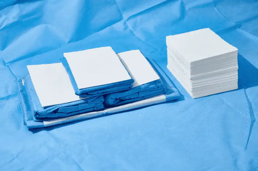
    Medical hand towels are specifically designed for use in healthcare environments. They are made from high-quality, absorbent materials that ensure quick and effective drying of hands while minimizing the spread of germs. The hygiene standards in healthcare settings demand a higher level of cleanliness, which is why using medical-grade towels is critical. These towels are typically more durable and are designed to withstand frequent use and washing, making them a reliable option for healthcare professionals.

    On the other hand, operating room paper towels are typically used in sterile environments where any potential contamination can pose a serious risk. These towels are specially designed to absorb moisture quickly while maintaining their integrity during surgical procedures. Due to the sterile nature of operating rooms, disposable paper towels are often preferred in such settings because they reduce the risk of infection and contamination.


    Why Choose Disposable Paper Towels?

    For many healthcare facilities, especially in high-traffic areas like emergency rooms and waiting areas, disposable paper towels offer an efficient and cost-effective solution. These towels are single-use, which significantly reduces the risk of cross-contamination. As an added benefit, disposable towels are easy to store, require no laundry, and come in individually wrapped packages that can help maintain hygienic conditions in any healthcare setting.

    The convenience of disposable towels also extends to environments where traditional cloth towels may not be suitable due to concerns about bacteria retention or infection risks. Whether for cleaning hands between patient interactions or for wiping surfaces, disposable paper towels ensure a quick, sanitary solution.


    Why Telijie’s Medical Hand Towels Stand Out

    When it comes to medical-grade hand towels, Telijie offers exceptional products that meet the highest healthcare standards. Known for its top-quality materials and attention to detail, Telijie’s medical hand towels are crafted to offer maximum absorbency, durability, and hygiene. These towels are rigorously tested to ensure that they meet the specific needs of medical professionals, providing both reliability and safety.

    Moreover, Telijie goes beyond just offering quality products. Their commitment to excellent customer service means that every healthcare facility, regardless of size, gets personalized attention and support. Whether you’re looking for operating room paper towels or disposable options, Telijie ensures a seamless purchasing experience and timely delivery, so you can focus on what matters most: patient care.


    In addition to product excellence, Telijie understands the unique needs of the healthcare industry and strives to build long-lasting relationships with its clients. Their customer service team is always available to answer any questions and provide guidance on the best towel options for your specific facility.

    In a world where hygiene is paramount, Telijie’s Medical hand towels offer the perfect balance of quality, convenience, and reliability. Whether you’re in need of operating room paper towels or prefer the ease of disposable paper towels, Telijie’s products ensure a cleaner, safer healthcare environment. Trust Telijie to provide not only top-tier medical hand towels but also unparalleled customer service to meet all your healthcare facility needs.

  • About SARS-CoV-2 (COVID-19) testing

    COVID-19 is an emerging infection and there is still much to learn about the mechanism about Covid-19 measurement. Nanjing Norman Biological Covid-19 solution, providing different Covid-19 testing kits to measure the antigen or antibody incurred by covid-19 is now available.


    The information these tests provide is essential to monitor and control the infection, and the development of effective therapeutic treatment and vaccines.


    Coronavirus Antigen Test Kit

    Time Kinetics of Antigen and Antibody Response in SARS-CoV-2

    Time kinetics of antibody response in coronavirus disease 2019 (COVID-19). The illustration demonstrates the relative levels of ARS-CoV-2 Antigen and host immunoglobulins (IgM, IgG) at different stages of COVID-19.


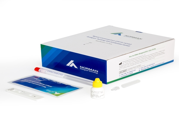
    Why choose Norman Novel Coronavirus (2019-nCoV) Antigen Testing Kit (Colloidal Gold)


    Virus detection in the laboratory is performed on nasopharyngeal and throat swabs using molecular PCR tests. PCR is the most sensitive method for detecting the virus early upon infection, sometimes before the onset of symptoms, and requires the use of specialized analyzers in a laboratory, often taking several hours to run the test.


    From a patient’s point of view, rapid antigen testing works in much the same way as molecular testing. Your health care provider will swab the back of your nose or throat to collect a sample for testing. But instead of waiting days for your results, an antigen test can produce a result in an hour or less, says the FDA.


    Norman biological COVID-19 Antigen kits are designed for the rapid detection of SARS-CoV-2 antigen in nasopharyngeal and throat swabs OR the saliva,final result takes 15mins .

    covid-19 rapid test kit


    These kits are for professional use and are intended as a screening test to aid in the early diagnosis of SARS-CoV-2 infection in patients with clinical symptoms. Positive results should be considered in conjunction with the clinical history and other data available to the clinician.


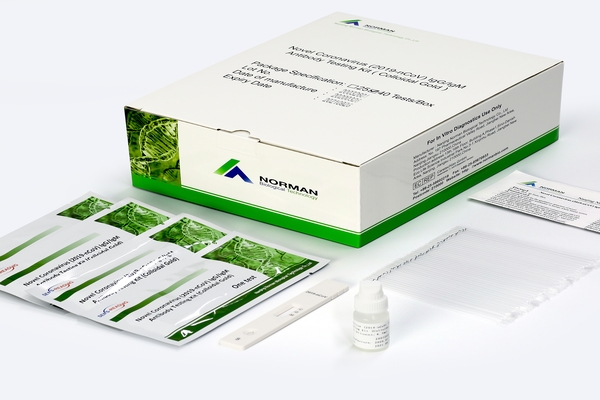
    Why Novel Coronavirus (2019-nCoV) IgG/IgM Antibody Testing Kit (Colloidal Gold)


    The detection of antibodies to SARS-Cov-2 in whole blood, serum or plasma samples is an indication of previous infection with the virus. As information about the antibody response to this virus increases, the clinical utility of laboratory and point of care tests will become clearer.

    Serological testing is a useful tool in the surveillance of the infection and in determining the extent of the COVID-19 pandemic. It may also be helpful for the diagnosis of patients with a negative PCR result or for the identification of patients that have had asymptomatic infections.

    COVID-19 IgG/IgM Rapid Test Kit

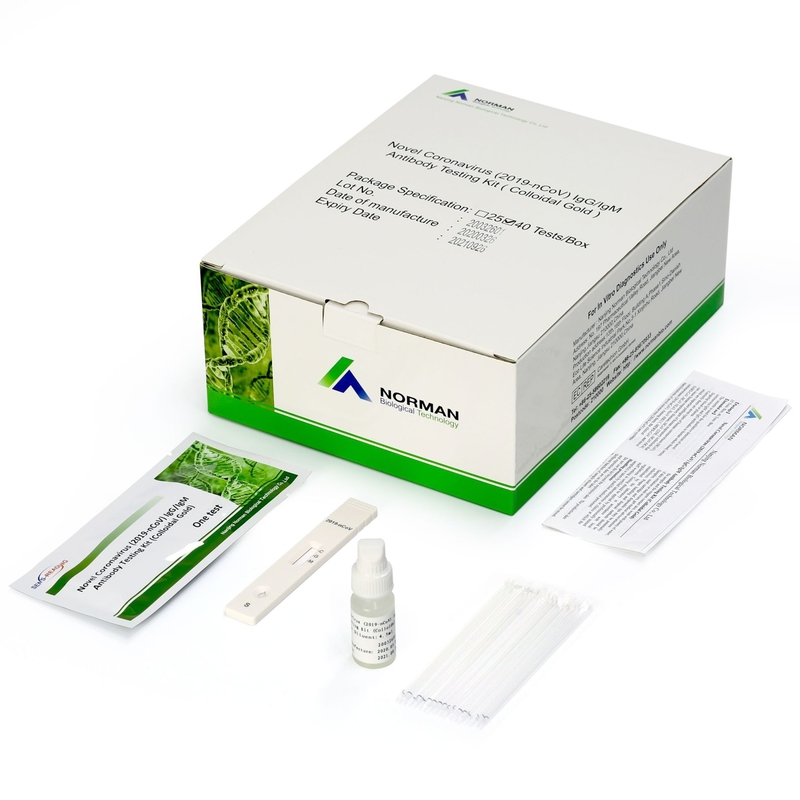
    Norman biological COVID-19 IgM/IgG Antibody rapid test is available for point of care antibody detection in whole blood from a finger-prick sample. These kits are not designed for home use and are available for professional use only in the global base. The results should be interpreted in conjunction with the clinical history of the individual, any previous exposure to the infection and the onset of any relevant symptoms. It has been shown that seroconversion rates are variable and it is recommended that samples for antibody detection are collected at least 2-3 weeks after onset of symptoms.

    Get more information from www.normanbio.com.


    Data sheet:

    https://www.normanbio.com/js/htmledit/kindeditor/attached/20210127/20210127165753_83816.pdf

    https://www.normanbio.com/js/htmledit/kindeditor/attached/20210116/20210116105841_49888.pdf

    https://www.normanbio.com/js/htmledit/kindeditor/attached/20210302/20210302160245_14732.pdf

    https://www.normanbio.com/js/htmledit/kindeditor/attached/20210302/20210302160353_87734.pdf


  • Neutralizing antibodies – the immune system's Superhero

    Neutralizing antibodies – the immune system’s Hero


    By Sebastian Fiedler, Lead Application Scientist Life Sciences at Fluidic Analytics


    Infectious diseases—a problem of the past or the present?

    Modern medicine has provided an exceptional set of tools to prevent and treat infectious diseases – conditions that used to decimate the human population with frightening regularity. As the world struggles to battle the COVID-19 pandemic, however, we are starkly reminded that infectious diseases are not merely a problem of the past.

    It is not only global pandemics like COVID-19 that evade the tools of modern medicine. We are in fact fighting this battle every day and on multiple fronts.  Humans remain under constant attack by bacteria and viruses, and even in modern times these pathogens still cause the death of millions of people around the world each year.

    How do you catch an infectious disease?

    Infectious diseases are caused by a variety of pathogenic microorganisms including bacteria, viruses, fungi and parasites. These pathogens typically enter our bodies through our mouths, eyes, noses or open wounds.

    Once inside our bodies, these microorganisms take advantage of their new favorable environment and quickly start multiplying. This process can severely damage and even kill the host cells resulting in visible and often well-described symptoms of specific diseases.

    Luckily, our bodies are not wholly without defense because the pathogens trigger an immune response to help fight the infection. This activity unfortunately can also create collateral damage within the body, with fever, rash, inflammation, and general malaise all hallmarks of an active immune response.

    Thankfully, modern medicine has delivered powerful antibiotics that are effective at supporting our own immune response across a broad spectrum of bacterial pathogens. The treatment of viral infections, however, has been much more challenging, and reliable therapeutics have so far eluded our best efforts. The most effective approach to fighting viral infections still is prevention by using vaccines that prime the immune system prior to a first encounter.


    What happens when a virus enters the body?

    When a virus enters the human body, it literally “goes viral”, producing as many copies of itself as possible. To do so, the virus exploits a cell’s own metabolism to release the new virus copies into the body, initiating the infection cycle over and over again. Once cells are infected, their natural function is badly impaired and, even worse, they often die. The resulting deficit of functional cells is the cause of tissue and organ failure, sometimes to an extent that is fatal.

    But as mentioned earlier, our immune system does not leave us defenseless against viruses. To prevent a large-scale viral spread in our bodies, several innate mechanisms protect us at each stage of infection.

    The first time we encounter a novel virus it typically avoids detection by the immune system and is able to enter a healthy cell.

    At this stage, this now-infected cell will utilize its internal defense mechanism to display fragments of the virus on its surface using special receptor proteins. This display of virus fragments alerts the body that the cell is infected and activates the immune system to kill and eliminate the cell before the virus can spread.

    In addition, the infected cells will also produce molecules, called interferons, which directly interfere with the process of viral replication to slow down the reproduction rate. Interferons also send a handy warning signal to nearby cells to alert them of the growing viral threat.

    Antibodies – our best defense

    The best defense against viruses, however, is to stop the infection in its tracks. This immune mechanism is made possible not by the cells themselves, but by antibodies which can identify and eliminate viruses before they start the infection cycle.

    Over the course of our lives, our bodies produce thousands of different types of antibodies that comprise our antibody-mediated immune response. Antibodies are proteins that are produced by B cells, which are a specialized type of blood cell. Once produced, these antibodies patrol our circulatory system and tissues, ready to deal with the pathogens.

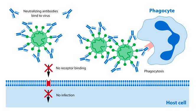
    Antibodies have several mechanisms to prevent infections. They can either neutralize viruses directly to prohibit their entry into the host cell, or they can crowd around a virus to increase its visibility to other immune cells. Once bound to a virus, antibodies can also tag the virus for phagocytes, which in turn ingest and destroy the pathogen.

    When a virus enters the human body, it literally “goes viral”,
    producing as many copies of itself as possible.

    Our antibodies’ ability to recognize and bind to pathogens starts with their structure. Fully assembled antibodies resemble the shape of the letter “Y”.

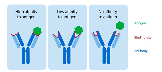
    The top of the two “arms” of the “Y” is where the magic happens. Imagine the arms as thousands of different jigsaw-puzzle pieces that give each antibody a unique shape. Each of those antibody jigsaw-puzzle pieces has the potential to fit specific virus antigens while fitting poorly with others.

    The better the antibody and antigen fit, the higher their affinity to each other. In other words, the stronger they bind to each other the more effective the antibody is at preventing infection by the virus.

    Novel Coronavirus (COVID-19) Neutralizing Antibody Testing

    Figure 1: The better the antibody and antigen fit, the stronger they bind to each other the more effective the antibody is at preventing infection by the virus.

    How do neutralizing antibodies earn their superhero status?

    To infect, viruses must first enter a healthy cell. They accomplish cell entry by binding to receptor molecules on the surface of their host cells. For example, SARS-CoV-2 utilizes so-called spike proteins on its surface for initial cell binding. These spike proteins fit perfectly to the shape of a receptor protein (ACE-2 receptor) typically found on the surface of human lung cells. Once the virus spike protein binds to the receptors of the lung cells, the virus enters and begins to replicate.

    Virus-neutralizing antibodies are designed to interfere with this binding event. To prevent entry to lung cells, an effective neutralizing antibody resembles the jigsaw-puzzle shape mimicking the lung-cell receptor ACE-2. In fact, it displays an even better fit than the receptor itself, resulting in the virus surface becoming covered by antibodies. This in turn prevents the virus from entering the lung cells. Moreover, such antibody-covered viruses become very sticky and attract each other to form large virus clusters, which, unlike individual viruses, are more easily recognized by other immune cells.

    Novel Coronavirus (COVID-19) Neutralizing Antibody Testing

    Figure 2: Neutralizing antibodies bind to spike proteins on the surface of SARS-CoV-2 and prevent the virus from binding and entering the host cell. As each antibody can bind to two spike proteins from different viruses, the start forming virus clusters that can be better recognised and destroyed by phagocytes.

    If antibodies offer such great protection, then why do some people become severely ill with COVID-19?

    This raises the question as to why some patients experience mild symptoms of COVID-19 while others suffer severely, or even die from, an otherwise identical disease.

    One hypothesis is that the progression and manifestation of the disease depends on the ability and strength of the antibodies in our bodies to protect us. It is believed that an antibody will be an effective neutralizer only if it fits perfectly to the shape of the spike proteins and therefore binds strongly to the virus (i.e., it binds with a high affinity).

    If a patient’s immune system produces only lower-affinity antibodies (or even non-neutralizing antibodies that do not block the receptor binding site of the spike protein at all), cellular protection becomes compromised, even though the immune system might try to compensate by producing increasing amounts of these weak or non-neutralizing antibodies.


    So are there tests that assess if we have neutralizing antibodies?

    Although it is reasonably straightforward to determine the presence of antibodies in COVID-19 patients using standard but crude immunoassays such as ELISA tests, assessing the virus-neutralizing capacity of antibodies in patient serum still relies on a cell-based neutralization assays. Although these assays do assess whether serum antibodies have the ability to block replication of the virus, they have significant drawbacks. They often require live biological materials and strict biosafety regulations, and most importantly, they are slow.

    Because they rely on the growth of living cells in culture, cell-based neutralization assays take several days to complete. During this time, a patient’s infection continues to develop. A multi-day wait for clinically actionable information could lead to several days of isolation from loved ones and absence from work at best; or a severe deterioration of symptoms and death at worst.

    Another limitation of cell-based neutralization assays specifically impacts the development of vaccines and antibody-based treatments. Current cell-based neutralization assays cannot characterize and quantify specific antigen–antibody interactions, but rather provide a binary readout of whether or not neutralizing antibodies are present. Because this binary outcome does not provide insights in the mechanisms of action of vaccines and therapeutics, key information about the relative effectiveness of vaccines or antibody-based treatments could be missed.

    No-fuzz detection and characterization in hours, not days!

    COVID-19 with all its impact on people, societies and everyday life, is a stark reminder that there is an urgent need in modern medicine for a test that informs patients, clinicians and researchers—in hours, not days—about the presence and, more importantly, the quality of neutralizing antibodies in their blood samples.

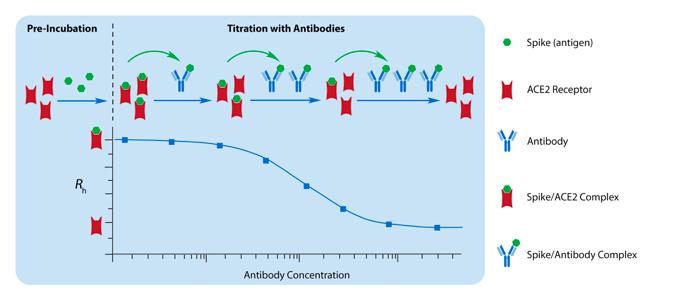
    To address this need, we have created a rapid, cell-free, virus-neuralization assay based on our in-solution technology and made it available on our Fluidity One-W Seruminstrument. The assay measures the binding interaction between the ACE-2 receptor and the SARS-CoV-2 spike protein, as well as the subsequent displacement of the spike protein in the presence of virus-neutralizing antibodies directly in patient serum, all within a couple of hours. Nanjing Norman Biological Technology Co.,Ltd

    Novel Coronavirus (COVID-19) Neutralizing Antibody Testing

    Figure 3. Our in-solution assay allows for the direct measurement of virus spike displacement from the ACE-2 receptor in the presence of neutralising antibodies.

    We are excited that this easy and fast approach could make a positive impact on patients’ lives and help researchers, clinicians and biopharma companies to better understand protective immunity and develop more effective vaccine and therapeutics candidates to fight COVID-19 (more information on this test shortly).

    Nanjing Norman Biological Technology Co.,Ltd is committing to offer comprehensive IVD solution covering self-developing and manufacturing raw material,reagent,equipment,get more COVID-19 Antibody Test Kit and Antigen Test Kit from https://www.normanbio.com.


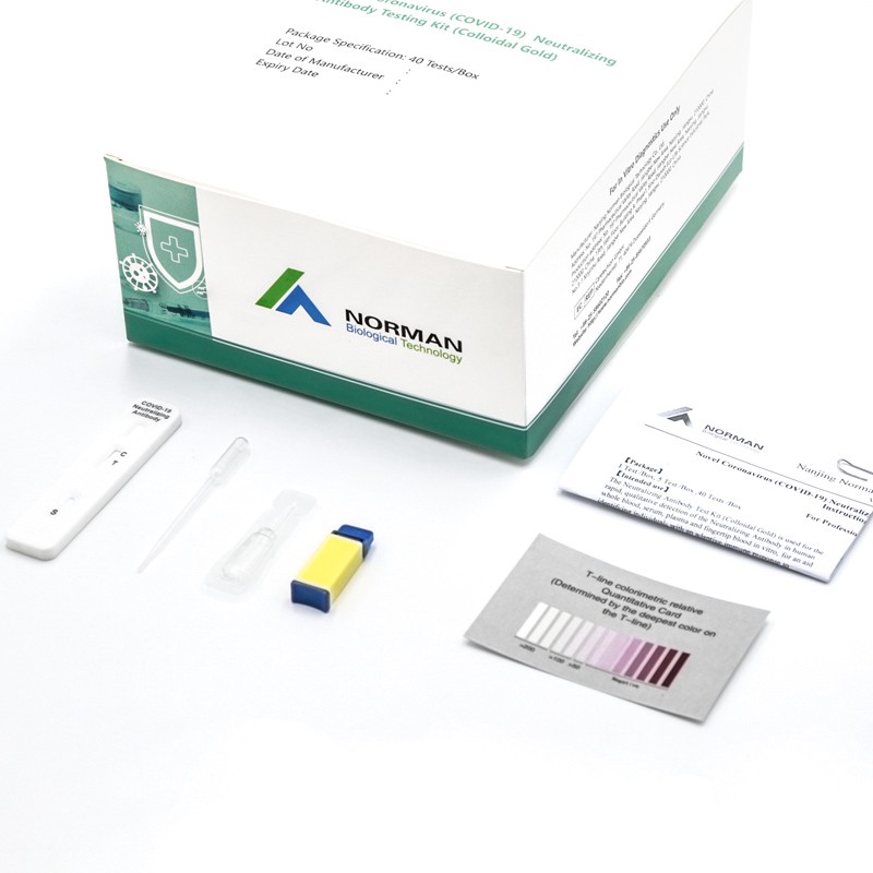
    Novel Coronavirus (COVID-19) Neutralizing Antibody Testing Kit (Colloidal Gold)


  • Saliva is more sensitive for SARS-CoV-2 detection in COVID-19 patients than nasopharyngeal swabs


    #SARS-CoV-2, COVID-19, saliva, diagnostics kit


    Abstract

    Rapid and accurate SARS-CoV-2 diagnostic testing is essential for controlling the ongoing COVID-19 pandemic. The current gold standard for COVID-19 diagnosis is real-time RT-PCR detection of SARS-CoV-2 from nasopharyngeal swabs. Low sensitivity, exposure risks to healthcare workers, and global shortages of swabs and personal protective equipment, however, necessitate the validation of new diagnostic approaches. Saliva is a promising candidate for SARS-CoV-2 diagnostics because (1) collection is minimally invasive and can reliably be self-administered and (2) saliva has exhibited comparable sensitivity to nasopharyngeal swabs in detection of other respiratory pathogens, including endemic human coronaviruses, in previous studies. To validate the use of saliva for SARS-CoV-2 detection, we tested nasopharyngeal and saliva samples from confirmed COVID-19 patients and self-collected samples from healthcare workers on COVID-19 wards. When we compared SARS-CoV-2 detection from patient-matched nasopharyngeal

    and saliva samples, we found that saliva yielded greater detection sensitivity and consistency throughout the course of infection. Furthermore, we report less variability in self-sample collection of saliva. Taken together, our findings demonstrate that saliva is a viable and more sensitive alternative to nasopharyngeal swabs and could enable at-home self-administered sample collection for accurate large-scale SARS-CoV-2 testing.

    Introduction

    Efforts to control SARS-CoV-2, the novel coronavirus causing COVID-19 pandemic, depend on accurate and rapid diagnostic testing. These tests must be (1) sensitive to mild and asymptomatic infections to promote effective self isolation and reduce transmission within high risk groups1 ; (2) consistent to reliably monitor disease progression and aid clinical decisions2 ; and (3) scalable to inform local and national public health policies, such as when social distancing measures can be safely relaxed. However, current SARS-CoV-2 testing strategies often fail to meet these criteria, in part because of their reliance on nasopharyngeal swabs as the widely recommended sample type for real-time RT-PCR.

    Although nasopharyngeal swabs are commonly used in respiratory virus diagnostics, they show relatively poor sensitivity for SARS-CoV-2 detection in early infection and are 2–6 inconsistent during serial testing . Moreover, collecting nasopharyngeal swabs causes discomfort to patients due to the procedure’s invasiveness, limiting compliance for repeat testing, and presents a considerable risk to healthcare workers, because it can induce patients to sneeze or cough, expelling virus particles7 . The procedure is also not conducive to large-scale testing, because there are widespread shortages of swabs and personal protective equipment for healthcare workers8 , and self-collection of nasopharyngeal swabs is difficult and less sensitive for virus detection9 . These challenges will be further exacerbated as the COVID-19 pandemic intensifies in low income countries. Given the limitations, a more reliable and less resource-intensive sample collection method, ideally one that accommodates self-collection in the home, is urgently needed. Saliva sampling is an appealing alternative to nasopharyngeal swab, since collecting saliva is non-invasive and easy to self-administer. An analysis of nasopharyngeal and saliva concordance for RT-PCR detection of respiratory pathogens, including two seasonal

    human coronaviruses, suggests comparable diagnostic sensitivity between the two sample 10,11of COVID-19 patients and (2) self-collected saliva samples have comparable. Preliminary findings indicate that (1) SARS-CoV-2 can be detected from the saliva 12 typesmSARS-CoV-2 detection sensitivity to nasopharyngeal swabs collected by healthcare13 workers from mild and subclinical COVID-19 cases . Critically, however, no rigorous evaluation of the sensitivity of SARS-CoV-2 detection in saliva with respect to nasopharyngeal swabs has been conducted from inpatients during the course of COVID-19 infection.

    In this study, we evaluated SARS-CoV-2 detection in paired nasopharyngeal swabs and saliva samples collected from COVID-19 inpatients and asymptomatic healthcare workers at moderate-to-high risk of COVID-19 exposure. Our results indicate that using saliva for SARS-CoV-2 detection is more sensitive and consistent than using nasopharyngeal swabs. Overall, we demonstrate that saliva should be considered as a reliable sample type to alleviate COVID-19 testing demands.

    Results

    Higher SARS-CoV-2 titers detected from saliva than nasopharyngeal swabs from inpatients
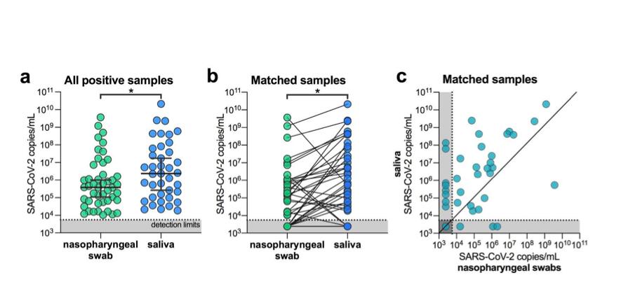
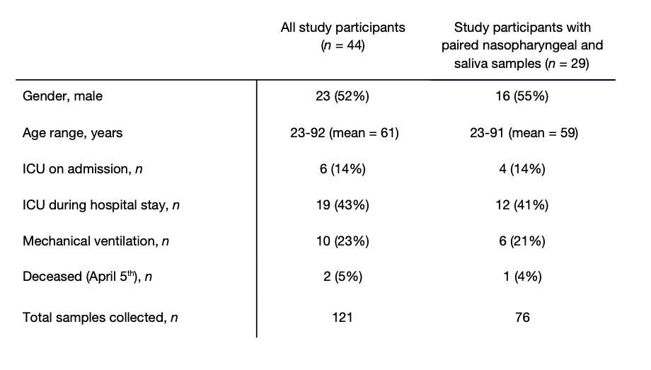
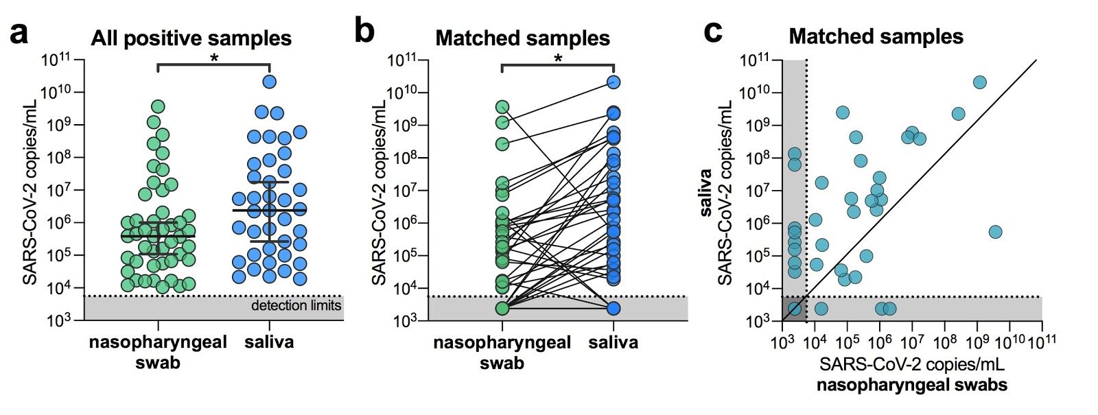
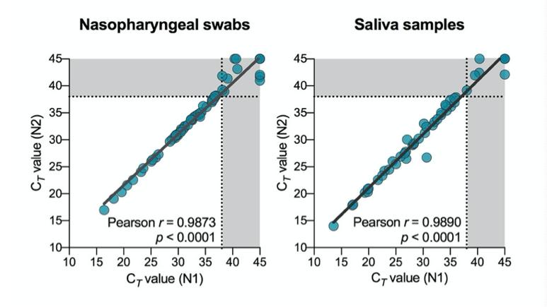
    To determine if saliva performs as well as the U.S. CDC recommendation of using nasopharyngeal swabs for SARS-CoV-2 diagnostics, we collected clinical samples from 44 COVID-19 inpatient study participants (Table 1). This cohort represents a range of COVID-19 patients with severe disease, with 19 (43%) requiring intensive care, 10 (23%) requiring mechanical ventilation, and 2 (5%) deceased as of April 5th, 2020. Using the U.S. CDC SARS-CoV-2 RT-PCR assay, we tested 121 self-collected saliva or healthcare worker-administered nasopharyngeal swabs from this cohort. We found strong concordance between the U.S. CDC “N1” and “N2” primer-probe sets (Extended Data Fig. 1), and thus calculated virus titers (virus copies/mL) using only the “N1” set. From all positive samples tested (n = 46 nasopharyngeal, 37 saliva), we found that the geometric mean virus titers from saliva were about 5⨉ higher than nasopharyngeal swabs (p < 0.05, Mann-Whitney test; Fig. 1a). When limiting our analysis to only patient-matched nasopharyngeal and saliva samples (n = 38 for each sample type), we found that SARS-CoV-2 titers from saliva were significantly higher than nasopharyngeal swabs (p = 0.0001, Wilcoxon test; Fig. 1b). Moreover, we detected SARS-CoV-2 from the saliva but not the nasopharyngeal swabs from eight matching samples (21%), while we only detected SARS-CoV-2 from nasopharyngeal swabs and not saliva from three matched samples (8%; Fig. 1c). Overall, we found higher SARS-CoV-2 titers from saliva than nasopharyngeal swabs from hospital inpatients.

    Table 1. COVID-19 inpatient cohort characteristics
    SARS-CoV-2 diagnostic testing

    SARS-CoV-2 diagnostic testing


    Figure 1. SARS-CoV-2 titers are higher in the saliva than nasopharyngeal swabs from hospital inpatients. (a) All positive nasopharyngeal swabs (n = 46) and saliva samples (n = 39) were compared by a Mann-Whitney test (p < 0.05). Bars represent the median and 95% CI. Our assay detection limits for SARS-CoV-2 using the US CDC “N1” assay is at cycle threshold 38, which corresponds to 5,610 virus copies/mL of sample (shown as dotted line and grey area). (b) Patient matched samples (n = 38), represented by the connecting lines, were compared by a Wilcoxon test test (p < 0.05). (c) Patient matched samples (n = 38) are also represented on a scatter plot. All of the data used to generate this figure, including the raw cycle thresholds, can be found in Supplementary Data 1. Extended Data Fig. 1 shows the correlation between US CDC assay “N1” and “N2” results.

    Less temporal SARS-CoV-2 variability when testing saliva from inpatients
    SARS-CoV-2 diagnostic testing

    As temporal SARS-CoV-2 diagnostic testing from nasopharyngeal swabs is reported to be 2,3 variable , we tested longitudinal nasopharyngeal and saliva samples from inpatients to determine which sample type provided more consistent results. From 22 participants with multiple nasopharyngeal swabs and 12 participants with multiple saliva samples, we found that SARS-CoV-2 titers generally decreased in both sample types following the reported date of symptom onset (Fig. 2a). Our nasopharyngeal swab results are consistent with 2,3 previous reports of variable SARS-CoV-2 titers and results : we found 5 instances where a participant’s nasopharyngeal swab was negative for SARS-CoV-2 followed by a positive result during the next collection (5/33 repeats, 33%; Fig. 2b) . In longitudinal saliva collections from 12 patients, however, there were no instances in which a sample tested negative and was later followed by a positive result. As true negative test results are important for clinicians to track patient improvements and for decisions regarding discharges, our data suggests that saliva is a more consistent sample type than nasopharyngeal swabs for monitoring temporal changes in SARS-CoV-2 titers.
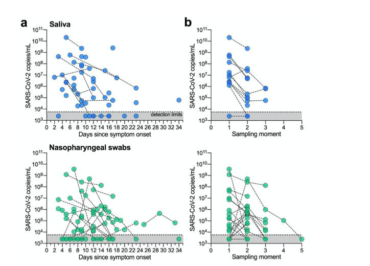
    Figure 2: SARS-CoV-2 detection is less variable between repeat sample collections with saliva. (a) Longitudinal SARS-CoV-2 titers from saliva or nasopharyngeal swabs are shown as days since symptom onset. Each circle represents a separate sample, which are connected to additional samples from the same patient by a dashed line. Our assay detection limits for SARS-CoV-2 using the US CDC “N1” assay is at cycle threshold 38, which corresponds to 5,610 virus copies/mL of sample (shown as dotted line and grey area). (b) The data are also shown by sampling moment (sequential collection time) to highlight the differences in virus titers between collection points. All of the data used to generate this figure, including the raw cycle thresholds, can be found in Supplementary Data 1.

    More consistent self-sampling from healthcare workers using saliva

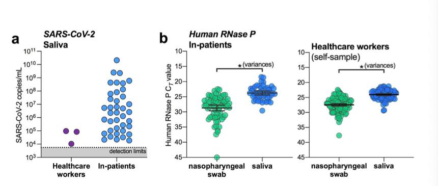
    Validating saliva for the detection of subclinical SARS-CoV-2 infections could prove transformative for both remote patient diagnostics and healthcare worker surveillance. To investigate this, we enrolled 98 asymptomatic healthcare workers into our study and collected saliva and/or nasopharyngeal swabs on average every 2.9 days (range = 1-8 days, Table 2). To date, we have detected SARS-CoV-2 in saliva from two healthcare workers who were negative by nasopharyngeal swabs using both the US CDC “N1” and “N2” tests and did not report any symptoms. The saliva from one of these individuals again tested positive alongside a matching negative nasopharyngeal swab upon repeat testing 2 days later. Virus titers from asymptomatic healthcare workers’ saliva are lower than what we typically detect from symptomatic inpatients (Fig. 3a), which likely supports the lack of symptoms.

    Our limited data supports that saliva may be more sensitive for detecting asymptomatic or pre-symptomatic infections; however, a larger sample size is needed to confirm. As nasopharyngeal swab sampling inconsistency may be one of the potential issues for false negatives (Fig. 2), monitoring an internal control for proper sample collection, human RNase P, may provide an alternative evaluation technique. While human RNase P detection was better from saliva in both the inpatient and healthcare worker cohorts (Fig. 3b) , this alone may not indicate better virus detection. More importantly, we found that human RNase P detection was more variable from nasopharyngeal swabs collected from inpatients (p = 0.0001, F test for variances) and self-collected from healthcare workers (p = 0.0002; Fig. 3b). Our results suggest that saliva may also be an appropriate, and perhaps more sensitive, alternative to nasopharyngeal swabs for screening asymptomatic or pre-symptomatic SARS-CoV-2 infections.

    Table 2. Healthcare worker cohort

    Figure 3. Saliva is an alternative for SARS-CoV-2 screening from healthcare workers and asymptomatic cases. (a) SARS-CoV-2 titers measured from the saliva of healthcare workers and inpatients. Our assay detection limits for SARS-CoV-2 using the US CDC “N1” assay is at cycle threshold 38, which corresponds to 5,610 virus copies/mL of sample (shown as dotted line and grey area). (b) RT-PCR cycle thresholds (Ct) values for human RNase P, and internal control for sample collection, from either inpatients (left panel) or health care workers (right panel) were compared by variances using the F test (p = 0.0001 for inpatients; p = 0.0002 for healthcare workers). All of the data used to generate this figure, including the raw cycle thresholds, can be found in Supplementary Data 1.

    Discussion

    Our study demonstrates that saliva is a viable and preferable alternative to nasopharyngeal swabs for SARS-CoV-2 detection. We found that the sensitivity of SARS-CoV-2 detection from saliva is comparable, if not superior to nasopharyngeal swabs in early hospitalization and is more consistent during extended hospitalization and recovery. Moreover, the detection of SARS-CoV-2 from the saliva of two asymptomatic healthcare workers despite negative matched nasopharyngeal swabs suggests that saliva may also be a viable alternative for identifying mild or subclinical infections. With further validation, widespread implementation of saliva sampling could be transformative for public health efforts: saliva self-collection negates the need for direct healthcare worker-patient interaction, a source of 14–16 several major testing bottlenecks and overall nosocomial infection risk , and alleviates supply demands on swabs and personal protective equipment.

    AsSARS-CoV-2viralloadsdifferbetweenmildandseverecases ,a limitation of our study is the primary focus on COVID-19 inpatients, many with severe disease. While more data are required to more rigorously compare the efficacy of saliva in the hospital setting to earlier in the course of infection, findings from two recent studies support its potential for 13,18 detecting SARS-CoV-2 from both asymptomatic individuals and outpatients . As 12 infectious virus has been detected from the saliva of COVID-19 patients , ascertaining the relationship between virus genome copies and infectious virus particles in the saliva of 19 pre-symptomatic individuals will play a key role in understanding the dynamics of 1,20 asymptomatic transmission.

    Stemming from the promising results for SARS-CoV-2 detection in asymptomatic 13

    individuals ,asalivaSARS-CoV-2detectionassayhasalreadygainedapprovalthroughthe 18

    U.S. Food and Drug Administration emergency use authorization . To meet the growing testing demands, however, our findings support the need for immediate validation and implementation of saliva for SARS-CoV-2 diagnostics in certified clinical laboratories.

    Methods

    Ethics

    All study participants were enrolled and sampled in accordance to the Yale University HIC-approved protocol #2000027690. Demographics, clinical data and samples were only collected after the study participant had acknowledged that they had understood the study protocol and signed the informed consent. All participant information and samples were collected in association with study identifiers.

    Participant enrollment

    Inpatients
    Patients admitted to Yale New Haven Hospital (a 1541-bed tertiary care medical center in New Haven, CT, USA), who tested positive for SARS-CoV-2 by nasopharyngeal and/or oropharyngeal swab (CDC approved assay) were invited to enroll in the research study. Exclusion criteria were age under 18 years, non-English speaking and clinical, radiological or laboratory evidence for a non-infectious cause of fever or respiratory symptoms or a microbiologically-confirmed infectious source (e.g. gastrointestinal, urinary, cardiovascular) other than respiratory tract for symptoms and no suspicion for COVID-19 infection.

    Healthcare workers
    Asymptomatic healthcare workers (e.g., without fever or respiratory symptoms) with occupational exposure to patients with COVID-19 were invited to enroll in the study. Study participation enabled active surveillance to ensure early detection following exposure and to further protect other healthcare workers and patients.

    Sample collection

    Inpatients
    Nasopharyngeal and saliva samples were obtained every three days throughout their clinical course. Nasopharyngeal samples were taken by registered nurses using the BD universal viral transport (UVT) system. The flexible, mini-tip swab was passed through the patient's nostril until the posterior nasopharynx was reached, left in place for several seconds to absorb secretions then slowly removed while rotating. The swab was placed in the sterile viral transport media (total volume 3 mL) and sealed securely. Saliva samples were self-collected by the patient. Upon waking, patients were asked to avoid food, water and brushing of teeth until the sample was collected. Patients were asked to repeatedly spit into a sterile urine cup until roughly a third full of liquid (excluding bubbles), before securely closing it. All samples were stored at room temperature and transported to the research lab at the Yale School of Public Health within 5 hours of sample collection.

    Healthcare workers
    Healthcare workers were asked to collect a self-administered nasopharyngeal swab and a saliva sample every three days for a period of 2 weeks. Samples were stored at +4°C until being transported to the research lab.

    SARS-CoV-2 detection

    On arrival at the research lab, total nucleic acid was extracted from 300 μl of viral transport media from the nasopharyngeal swab or 300 μl of whole saliva using the MagMAX Viral/Pathogen Nucleic Acid Isolation kit (ThermoFisher Scientific) following the manufacturer's protocol and eluted into 75 μl of elution buffer. For SARS-CoV-2 RNA 21,22 detection, 5 μl of RNA template was tested as previously described , using the US CDC real-time RT-PCR primer/probe sets for 2019-nCoV_N1 and 2019-nCoV_N2 and the human RNase P (RP) as an extraction control. Samples were classified as positive for SARS-CoV-2 when both N1 and N2 primer-probe sets were detected <38 C . Virus copies were T quantified using a 10-fold dilution standard curve of RNA transcripts that we previously 21 generated . As results from N1 and N2 were comparable (Extended Data Fig. 1), all virus copies are shown as calculated using the N1 primer-probe set.

    Statistical analysis

    Statistical analyses were conducted in GraphPad Prism 8.0.0 as described in the Results.

    Acknowledgments

    We gratefully acknowledge the study participants for their time and commitment to the study. We thank all members of the clinical team at Yale-New Haven Hospital for their dedication and work which made this study possible. We also thank S. Taylor and P. Jack for technical discussions.

    Funding

    The study was partially funded by the Yale Institute for Global Health. The corresponding authors had full access to all data in the study and had final responsibility for the decision to submit for publication.

    Extended data


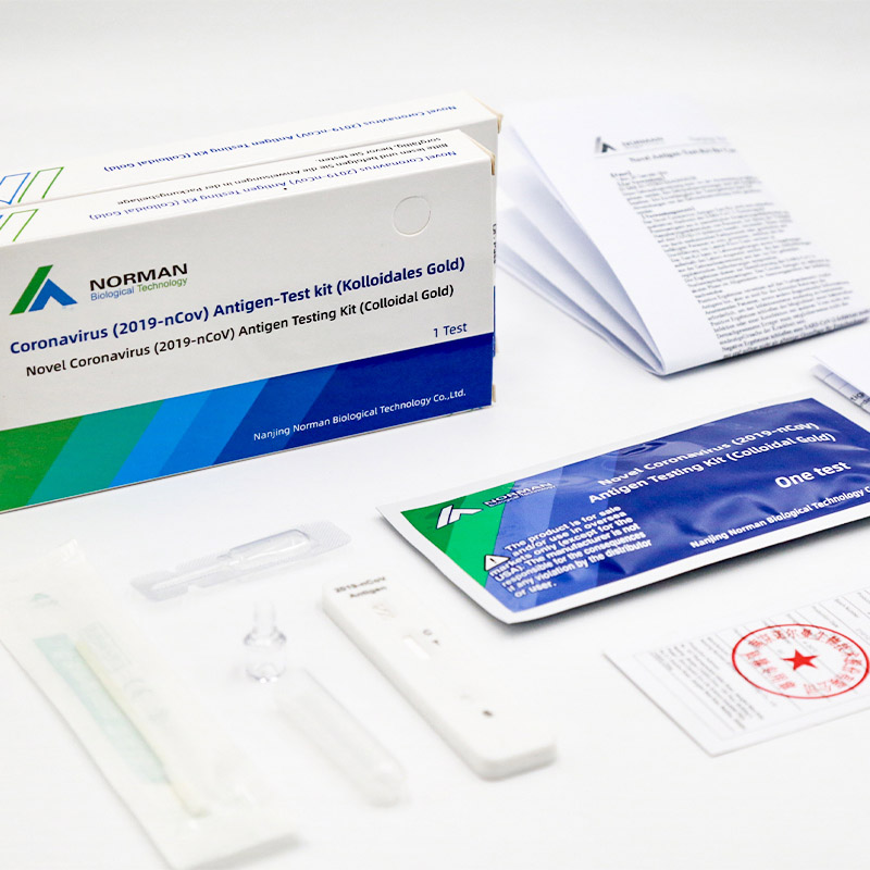
    Novel Coronavirus (2019-nCoV) Anterior Nasal Swab Antigen Testing Kit (Colloidal Gold)

    Novel Coronavirus (2019-nCoV) Anterior Nasal Swab Antigen Testing Kit (Colloidal Gold)

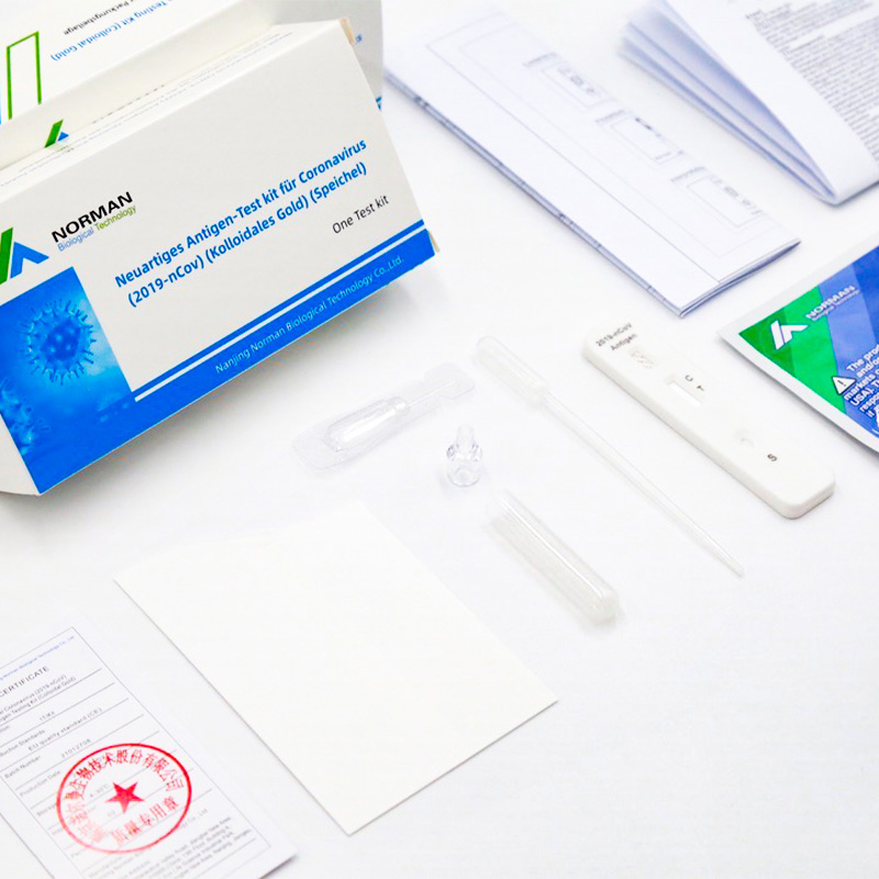
    Novel Coronavirus (2019-nCoV) Paper Saliva Antigen Testing Kit (Colloidal Gold)

    Novel Coronavirus (2019-nCoV) Paper Saliva Antigen Testing Kit (Colloidal Gold)


    Extended Data Fig. 1. Concordance between SARS-CoV-2 detection using US CDC “N1” and “N2” primer and probe sets. Ct = RT-PCR cycle threshold. Dotted line and grey areas indicate the limits of detection.

    References

    1. Kimball, A. et al. Asymptomatic and Presymptomatic SARS-CoV-2 Infections in Residents of a Long-Term Care Skilled Nursing Facility - King County, Washington, March 2020. MMWR Morb. Mortal. Wkly. Rep. 69, 377–381 (2020).

    2. Wölfel, R. et al. Virological assessment of hospitalized patients with COVID-2019. Nature (2020) doi:10.1038/s41586-020-2196-x.

    3. Zou, L. et al. SARS-CoV-2 Viral Load in Upper Respiratory Specimens of Infected Patients. N. Engl. J. Med. 382, 1177–1179 (2020).

    4. Zhao, J. et al. Antibody responses to SARS-CoV-2 in patients of novel coronavirus disease 2019. Clin. Infect. Dis. (2020) doi:10.1093/cid/ciaa344.

    5. Xie, X. et al. Chest CT for Typical 2019-nCoV Pneumonia: Relationship to Negative RT-PCR Testing. Radiology 200343 (2020).

    6. Wang, W. et al. Detection of SARS-CoV-2 in Different Types of Clinical Specimens. JAMA (2020) doi:10.1001/jama.2020.3786.

    7. To, K. K.-W. et al. Temporal profiles of viral load in posterior oropharyngeal saliva samples and serum antibody responses during infection by SARS-CoV-2: an observational cohort study. Lancet Infect. Dis. (2020) doi:10.1016/S1473-3099(20)30196-1.

    8. CDC. Interim Infection Prevention and Control Recommendations for Patients with Suspected or Confirmed Coronavirus Disease 2019 (COVID-19) in Healthcare Settings. Centers for Disease Control and Prevention https://www.cdc.gov/coronavirus/2019-ncov/hcp/infection-control-recommendations.h tml?CDC_AA_refVal=https%3A%2F%2Fwww.cdc.gov%2Fcoronavirus%2F2019-ncov %2Finfection-control%2Fcontrol-recommendations.html (2020).

    9. Dhiman, N. et al. Effectiveness of patient-collected swabs for influenza testing. Mayo Clin. Proc. 87, 548–554 (2012).

    10. Kim, Y.-G. et al. Comparison between Saliva and Nasopharyngeal Swab Specimens for Detection of Respiratory Viruses by Multiplex Reverse Transcription-PCR. J. Clin. Microbiol. 55, 226–233 (2017).

    11. Wyllie, A. L. et al. Molecular surveillance of nasopharyngeal carriage of Streptococcus pneumoniae in children vaccinated with conjugated polysaccharide pneumococcal vaccines. Sci. Rep. 6, 23809 (2016).

    12. To, K. K.-W. et al. Consistent Detection of 2019 Novel Coronavirus in Saliva. Clin. Infect. Dis. (2020) doi:10.1093/cid/ciaa149.

    13. Kojima, N. et al. Self-Collected Oral Fluid and Nasal Swabs Demonstrate Comparable Sensitivity to Clinician Collected Nasopharyngeal Swabs for Covid-19 Detection. medRxiv 2020.04.11.20062372 (2020).

    14. Tran, K., Cimon, K., Severn, M., Pessoa-Silva, C. L. & Conly, J. Aerosol generating procedures and risk of transmission of acute respiratory infections to healthcare workers: a systematic review. PLoS One 7, e35797 (2012).

    15. Judson, S. D. & Munster, V. J. Nosocomial Transmission of Emerging Viruses via Aerosol-Generating Medical Procedures. Viruses 11, (2019).

    16. Wang, D. et al. Clinical Characteristics of 138 Hospitalized Patients With 2019 Novel Coronavirus-Infected Pneumonia in Wuhan, China. JAMA (2020) doi:10.1001/jama.2020.1585.

    17. Liu, Y. et al. Viral dynamics in mild and severe cases of COVID-19. Lancet Infect. Dis. (2020) doi:10.1016/S1473-3099(20)30232-2.

    18. U.S. Food & Drug Administration. Accelerated Emergency Use Authorization (EUA) Summary SARS-CoV-2 Assay (Rutgers Clinical Genomics Laboratory). https://www.fda.gov/media/136875/download.

    19. Lauer, S. A. et al. The Incubation Period of Coronavirus Disease 2019 (COVID-19) From Publicly Reported Confirmed Cases: Estimation and Application. Ann. Intern. Med.

    Source: https://www.medrxiv.org/content/10.1101/2020.04.16.20067835v1.full.pdf


    AnneL.Wyllie1*,JohnFournier2, ArnauCasanovas-Massana1, MelissaCampbell2, MariaTokuyama3, Pavithra Vijayakumar4 , Bertie Geng4 , M. Catherine Muenker1 , Adam J. Moore1 , Chantal B.F. Vogels1 , Mary E. Petrone1 , Isabel M. Ott5, Peiwen Lu3 , Arvind Venkataraman3 , Alice Lu-Culligan3 , Jonathan Klein3 , Rebecca Earnest1 , Michael Simonov6 , Rupak Datta2 , Ryan Handoko2 , Nida Naushad2 , Lorenzo R. Sewanan2 , Jordan Valdez2 , Elizabeth B. White1 , Sarah Lapidus1 , Chaney C. Kalinich1 , Xiaodong Jiang3 , Daniel J. Kim3 , Eriko Kudo3 , Melissa Linehan3 , Tianyang Mao3 , Miyu Moriyama3 , Ji Eun Oh3 , Annsea Park3 , Julio Silva3 , Eric Song3 , Takehiro Takahashi3 , Manabu Taura3 , Orr-El Weizman3 , Patrick Wong3 , Yexin Yang3 , Santos Bermejo7 , Camila Odio8 , Saad B. Omer1,2,9,10, Charles S. Dela Cruz7 , ShelliFarhadian2, RichardA.Martinello2,7,11, AkikoIwasaki3,12, NathanD.Grubaugh1#*,AlbertI.Ko1#*

    1 Department of Epidemiology of Microbial Diseases, Yale School of Public Health, New Haven, CT 06510, USA 2 Department of Medicine, Section of Infectious Diseases, Yale School of Medicine, New Haven, CT, 06510, USA
    3 Department of Immunobiology, Yale School of Medicine, New Haven, CT, 06510, USA
    4 Department of Obstetrics, Gynecology, and Reproductive Sciences, Yale School of Medicine, New Haven, CT, 06510, USA
    5 Department of Ecology and Evolutionary Biology, Yale University, New Haven, CT 06520, USA
    6 Program of Applied Translational Research, Yale School of Medicine, New Haven, CT, 06510, USA
    7 Department of Internal Medicine, Section of Pulmonary, Critical Care, and Sleep Medicine, Yale School of Medicine, New Haven, CT, 06510, USA
    8 Department of Medicine, Northeast Medical Group, Yale-New Haven Health, New Haven, CT 06510, USA
    9 Yale Institute of Global Health, New Haven, CT 06510, USA
    10 Yale School of Nursing, New Haven, CT 06510, USA
    11 Department of Infection Prevention, Yale-New Haven Health, New Haven, CT 06520
    12 Howard Hughes Medical Institute, New Haven, CT 06510, USA

    # Joint senior authors
    * Correspondence: anne.wyllie@yale.edu (ALW); nathan.grubaugh@yale.edu (NDG); albert.ko@yale.edu (AIK)


  • The Principle of Direct Chemiluminescence Immunoassay

          Chemiluminescence Analyzer consists of the two parts of fully automated reaction system and chemiluminescence assay system. Fully automated reaction system is the process of indirect specific binding of luminescent marker to solid phase carrier by means of incubation and magnetic separation. Chemiluminescence assay system refers to the process of photomultiplier’s capture of photon with maximum wavelength of 430nm emitted by luminescent marker upon oxidization with hydrogen peroxide when acid environment suddenly changes into alkaline environment.

          The system uses acridinium ester and its derivatives as luminescent marker, and it becomes luminous through injection of activator within seconds, which makes it flash-type chemiluminescence. As luminescent marker used for assay and analysis, acridinium ester and its derivatives boasts of simple reaction, rapidness, absence of catalyst, excellent stability, less nonspecific binding, high sensitivity and other strengths, which make it a new effective compound.

          The core detector of this instrument is photomultiplier (PMT), which detects single photons and transfer to amplifier. High-voltage current’s added for amplification, amplifier converts analog current into digital current, digital current transfer luminescence signal to mainboard for calculation of relative luminescence unit (RLU), and contents of test antigens or antibodies in measurement sample are calculated through standard curve.

  • Understanding the Chemiluminescence Test Applications and Importance

    Chemiluminescence tests are a fascinating intersection of chemistry and biology, widely utilized in scientific research and practical applications. But what exactly is this test, and why is it so valuable? Lets dive into its principles, uses, and significance.
    What is Chemiluminescence?
    Chemiluminescence refers to the emission of light during a chemical reaction without the involvement of external light sources like fluorescence or phosphorescence. This process occurs when an excited intermediate molecule releases energy in the form of visible or near-visible light as it returns to its ground state.
    In tests, chemiluminescence is harnessed to detect and measure specific substances, as the emitted light is directly proportional to the concentration of the target compound.
    How Does the Chemiluminescence Test Work?
    1. Reaction Setup: A sample containing the substance of interest is mixed with reagents that trigger the chemiluminescent reaction.
    2. Light Emission: The reaction produces light, often with the help of enhancers or catalysts.
    3. Detection: Instruments like luminometers or specialized cameras capture the emitted light, quantifying the substance of interest based on its intensity.
    Key Applications of the Chemiluminescence Test
    1. Medical Diagnostics
    Chemiluminescence immunoassays (CLIA) are widely used in medical testing. These tests detect hormones, proteins, or antigens in blood or other bodily fluids, aiding in diagnosing diseases such as cancer, autoimmune disorders, and infectious diseases.
    2. Environmental Monitoring
    Chemiluminescence plays a role in detecting pollutants like nitrogen oxides (NOx) in the atmosphere. This application is critical for understanding air quality and mitigating environmental hazards.
    3. Pharmaceutical and Food Industries
    These tests help ensure quality control by detecting contaminants or verifying the concentration of active compounds in drugs and food products.
    4. Forensic Science
    In forensic investigations, chemiluminescence is used to detect trace amounts of substances, such as blood at crime scenes. Luminol, a chemiluminescent reagent, is a classic example used for such purposes.
    5. Biological Research
    Researchers use chemiluminescence to study cellular processes, gene expression, or protein interactions. It is a sensitive method for tracking biomolecular activity.
    Advantages of Chemiluminescence Tests
    - High Sensitivity: Detects even trace amounts of substances.
    - Specificity: Reactions can be tailored for specific targets.
    - Rapid Results: Quick reaction times lead to fast readings.
    - Low Background Noise: Absence of excitation light reduces interference.
    Challenges and Limitations
    While powerful, chemiluminescence tests require precise conditions for optimal performance. Factors like reagent stability, light measurement accuracy, and the presence of interfering substances can influence results.
    Conclusion
    The chemiluminescence test is a versatile tool that spans disciplines, from healthcare to environmental science. Its ability to provide accurate and rapid analysis makes it indispensable in modern science and industry. Whether used to detect diseases, pollutants, or forensic evidence, this test continues to illuminate pathways to innovation and discovery.
    What do you think about the applications of chemiluminescence in your field? Let us know in the comments!

  • Why Choose Antibody Test ?

    Why Choose Antibody Test

    Although the English expert from the World Health Organization (WHO) subsequently tested negative, it was not immediately clear if the earlier result was a false positive, or the result of previous infection or a Sars-CoV-2 vaccination.


    WILL VACCINATED PEOPLE GET POSITIVE ANTIBODY RESULTS?

    It is possible, but not always, experts say. Most vaccines target the “spike” protein on the virus surface to trigger an immune response that could include IgM antibodies.

    “We can assume that any Sars-CoV-2 vaccine containing the spike protein will induce IgM and therefore a diagnostic assay designed to detect spike specific IgM will not be able to differentiate vaccination from infection,” said Helen Fletcher, a professor of immunology at the London School of Hygiene & Tropical Medicine.

    Published data on Oxford University/AstraZeneca Plc’s Sars-CoV-2 vaccine shows spike protein-triggered IgM is detectable in some people at least 56 days after immunization, Fletcher said.


    IS IT POSSIBLE TO USE DIFFERENT ANTIBODY TESTS?

    Tests to detect antibodies triggered by non-spike protein can yield negative results for those who got vaccines targeting spike protein, said Jin Dong-Yan, a professor of virology at the University of Hong Kong.

    Vaccines targeting spike protein include those from AstraZeneca, Pfizer Inc and its partner BioNTech, and Moderna.


    THERE ARE STILLNEED TO BE CLARIFIED

    Such tests, But, can be problematic for other types of vaccines, including whole virus-based shots .some experts said.

    “Where a person is injected with whole virus-based inactivated Sars-CoV-2 vaccine...there’s a strong chance that the person may also have positive result from non-spike protein IgM antibody tests,” said Ian Jones, a virologist at Britain’s University of Reading.


    Hot Products:

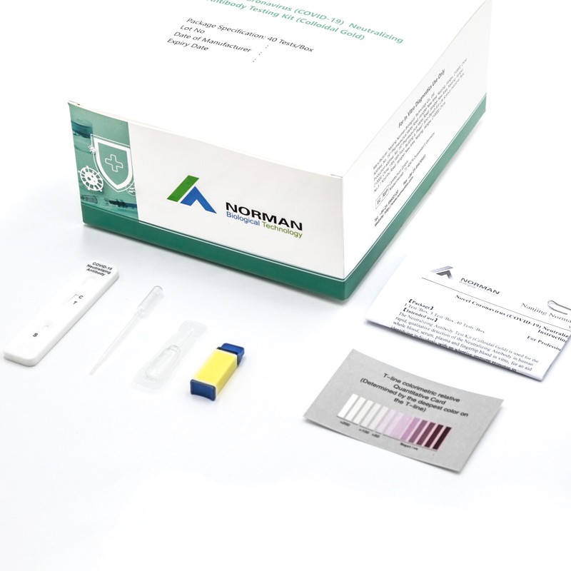
    COVID-19 Neutralizing Antibody Testing Kit


    COVID-19 Neutralizing Antibody Testing Kit


    Novel Coronavirus IgG/IgM Antibody Testing Kit


  • Why Do We Need To Take Care Of Our Hearts?

    Cardiovascular disease (CVD) is the world's number one killer, causing over 18.6 million deaths per year. CVD is a class of diseases that affect the heart or blood vessels (veins and arteries). More people die from CVD worldwide than from any other cause: over 18.6 million every year. Of these deaths, 85% are due to coronary heart diseases (e.g heart attacks) and cerebrovascular diseases (e.g. strokes) and mostly affect low-and middle-income countries.

    Do you know that sometimes medicines or irregularities in your heart rate may warrant a visit to your doctor? And, if your pulse is very low, very high, or frequently switches between, you also need to consult a doctor right away?

    Keeping track of your health and wellness has become more crucial than ever. Everything matters: what you eat, how much Sleep you get, are you working out, and what you are doing to keep your mind, body, and soul intact.

    Working from nine to five, looking after the family and yourself, can be overwhelming. But you cannot avoid them. You must take care of their heart.

    Maintaining good health will avoid problems like absenteeism or other physical, mental, emotional, and spiritual imbalance and remain productive. It will help you thrive in both your personal and professional life. So, setting your health goals today will help you succeed in the long run.

    Every heartbeat counts, and the health of every employee matters. Together let's care for our heart health and take action to beat cardiovascular disease.

    As a top in vitro diagnostic equipment enterprise in China, Normanbio has been at the forefront of developing comprehensive tests in aid of evaluating the risk of cardiovascular diseases, which enables healthcare professionals to provide real-time diagnosis, make informed treatment decisions and improve patient outcomes.


  • The Ultimate Solution for Clean Dining Disposable Bibs for Every Occasion

    Dining out or enjoying a family meal at home can often lead to messy situations, especially when it comes to young children or even adults who tend to spill food. This is where a disposable bib comes in handy. A disposable dining bib offers a convenient, hygienic, and practical solution to prevent stains and keep your clothing clean while enjoying your meal. Whether you're at a restaurant or at home, disposable bibs are becoming a must-have for any dining situation.

    The disposable bib is designed with simplicity and efficiency in mind. Made from high-quality, lightweight materials, these bibs provide comfort and durability while being easy to dispose of after use. Available in different sizes and styles, disposable dining bibs cater to various needs, from protecting your clothes during meals to ensuring cleanliness during messy activities like crafting or painting. These bibs are especially popular for children, who are often more prone to creating a mess when eating.

    One of the standout options in the market today is the disposable printing bib, which offers customization options for businesses and events. Whether you're looking to create personalized bibs for a restaurant, a promotional giveaway, or an event, these printed bibs can be branded with logos, images, or slogans, providing an extra touch of marketing while serving a functional purpose. The disposable printing bib can help enhance your business’s visibility, all while offering a clean and practical solution for your customers.

    At Telijie, we understand the importance of both product quality and service excellence. As a trusted manufacturer, we offer a range of high-quality disposable bib options, including disposable dining bibs and disposable printing bibs, that meet the highest standards of hygiene and comfort. Our products are designed to be gentle on the skin yet highly durable, ensuring that you can enjoy your meals without worry. Beyond the product itself, Telijie is committed to providing exceptional service to our clients. With efficient production timelines, reliable shipping, and customizable options, we ensure that your business or personal needs are met with utmost satisfaction.

    Whether you’re looking for a practical solution for your restaurant, a promotional item for your event, or a way to keep your family clean during mealtime, Telijie’s range of disposable bibs offers the perfect combination of convenience, style, and functionality. With our expertise in providing both quality products and top-tier service, you can trust Telijie to meet all your disposable bib needs with professionalism and efficiency.