• The Functions of insulin cooler bag

    |

    The function of an insulin bag is to provide a portable and insulated storage solution for insulin and other diabetes supplies. Insulin needs to be kept at a specific temperature range to maintain its effectiveness, usually between 2°C and 8°C (36°F and 46°F) or as specified by the manufacturer.

    An insulin bag is designed with insulation materials to help maintain the temperature stability of insulin during travel or daily routines. It helps protect insulin from extreme temperatures, whether it's hot or cold, which can negatively affect its potency. By keeping insulin within the recommended temperature range, an insulin bag helps ensure that it remains safe and effective for use in managing diabetes.

    Insulin bags are often equipped with cooling elements such as gel packs or ice packs that can be frozen and inserted into the bag to provide additional temperature control. Some insulin bags also come with temperature monitoring features, like built-in thermometers or digital displays, to allow users to monitor and adjust the temperature inside the bag.

    Overall, the primary function of an insulin bag is to provide a convenient and reliable way to store insulin while maintaining its temperature stability, allowing individuals with diabetes to carry their medication with them wherever they go.

  • What's The Principle of Immunofluorescence Assays?

    Immunofluorescence (IF) is a powerful approach for getting insight into cellular structures and processes using microscopy. Specific proteins can be assessed for their expression and location, making immunofluorescence indispensable for scientists to solve many cell biological questions.

    An immunofluorescence experiment is based on the following principal steps:


    1. Specific antibodies bind to the protein of interest.

    2. Fluorescent dyes are coupled to these immune complexes in order to visualize the protein of interest using microscopy.


    It is distinguished between direct and indirect immunofluorescence. In direct immunofluorescence, the primary antibody is directly coupled to a fluorophore (also called fluorochrome), allowing for easy handling and quick visualization. In indirect immunofluorescence, a secondary fluorophore-coupled antibody, which specifically binds to the primary antibody, is used to visualize the structure of interest.


    Although the second approach is more time-consuming than direct immunofluorescence, it has several big advantages, such as it is generally less expensive, because the secondary antibody can be used for different primary antibodies. In addition, several proteins can be specifically visualized in parallel in one single sample (multicolor immunofluorescence) by combining multiple primary antibodies with specific secondary antibodieseach of them labeled with a different fluorophore.


    Nanjing Norman is a complete innovative diagnostic enterprise integrating research and sales. It has a comprehensive technical platform with complete instruments, reagents and reagent raw materials.


    The company has successfully listed on the market with immunofluorescence analyzer FI-1000, FI-1200 and hundreds of testing items.


    Nanjing Norman will continue to provide higher-quality products and services to global customers and partners! Welcome to send inquiry to info@nrmchina.com


  • Unveiling the Precision of Chemiluminescence Immunoassay in Modern Diagnostics

    Welcome to our blog, where we delve into the fascinating realm of healthcare diagnostics and shine a light on one of the most innovative technologies in the field – Chemiluminescence Immunoassay (CLIA). In this article, we will unravel the mysteries behind CLIA, its applications, and the significant impact it has on modern medical diagnostics.  Normanbio is professional Chemiluminescence Immunoassay analyzer and diagnostic equipment manufacturer.


    What is Chemiluminescence Immunoassay (CLIA)?
    Chemiluminescence Immunoassay, or CLIA, is a powerful and highly sensitive laboratory technique used for detecting the presence of specific molecules in biological samples. It combines the principles of immunoassay with the unique property of chemiluminescence – the emission of light resulting from a chemical reaction.

    Key Components of CLIA:

    Antibody-Antigen Interaction: CLIA relies on the specificity of antibodies to selectively bind to target antigens. This interaction forms the basis of the assay's accuracy.

    Luminogenic Substrates: The magic happens when a luminogenic substrate is introduced. This substance reacts with an enzyme attached to the antibody-antigen complex, resulting in the emission of light.

    Photodetectors: Specialized instruments capture and measure the emitted light, providing quantitative data about the concentration of the target molecule in the sample.

    Applications of CLIA:

    Clinical Diagnostics: CLIA is widely employed in clinical laboratories to diagnose various medical conditions, including infectious diseases, hormonal disorders, and autoimmune diseases.

    Therapeutic Drug Monitoring: It plays a crucial role in monitoring drug levels in patients undergoing therapeutic treatments, ensuring optimal dosage and minimizing adverse effects.

    Research and Development: CLIA is a valuable tool in pharmaceutical research, aiding in the development and testing of new drugs and therapies.

    Advantages of CLIA:

    High Sensitivity and Specificity: CLIA offers exceptional sensitivity, enabling the detection of low concentrations of target molecules with high precision.

    Wide Dynamic Range: The technique can quantify a broad range of analyte concentrations, making it versatile for different diagnostic applications.

    Automation and Throughput: Automated CLIA systems enhance efficiency, enabling high-throughput analysis, making it suitable for large-scale testing.

    In conclusion, Chemiluminescence Immunoassay stands as a beacon of innovation in the field of diagnostics, illuminating new possibilities for accurate and efficient detection of various biomolecules. Its applications in clinical settings, research laboratories, and pharmaceutical development underscore its significance in advancing healthcare. As technology continues to evolve, CLIA is poised to play an even more prominent role in shaping the future of diagnostic medicine. Stay tuned for more updates on the latest advancements in healthcare technology on Norman's blog.
  • PFBN Proximal Femoral Bionic Intramedullary Nail Clinical Cases

    Intertrochanteric fracture of femur is one of the common hip fractures in the elderly. The key of treatment are the strong fixation and early rehabilitation exercise, which can effectively reduce the occurrence of complications caused by bed rest and is of great importance to improve the recovery. However, the failure of traditional fracture internal fixation system is common in the treatment process.  Recently, Liu Tao, director of the Department of Trauma and Orthopedics of Henan Provincial People's Hospital, took the lead in the first use of the international initiative and leading concept of the latest PFBN intramedullary nail fixation system in the treatment of intertrochanteric fracture successfully.

    Case Information:

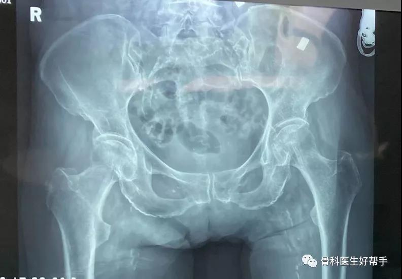
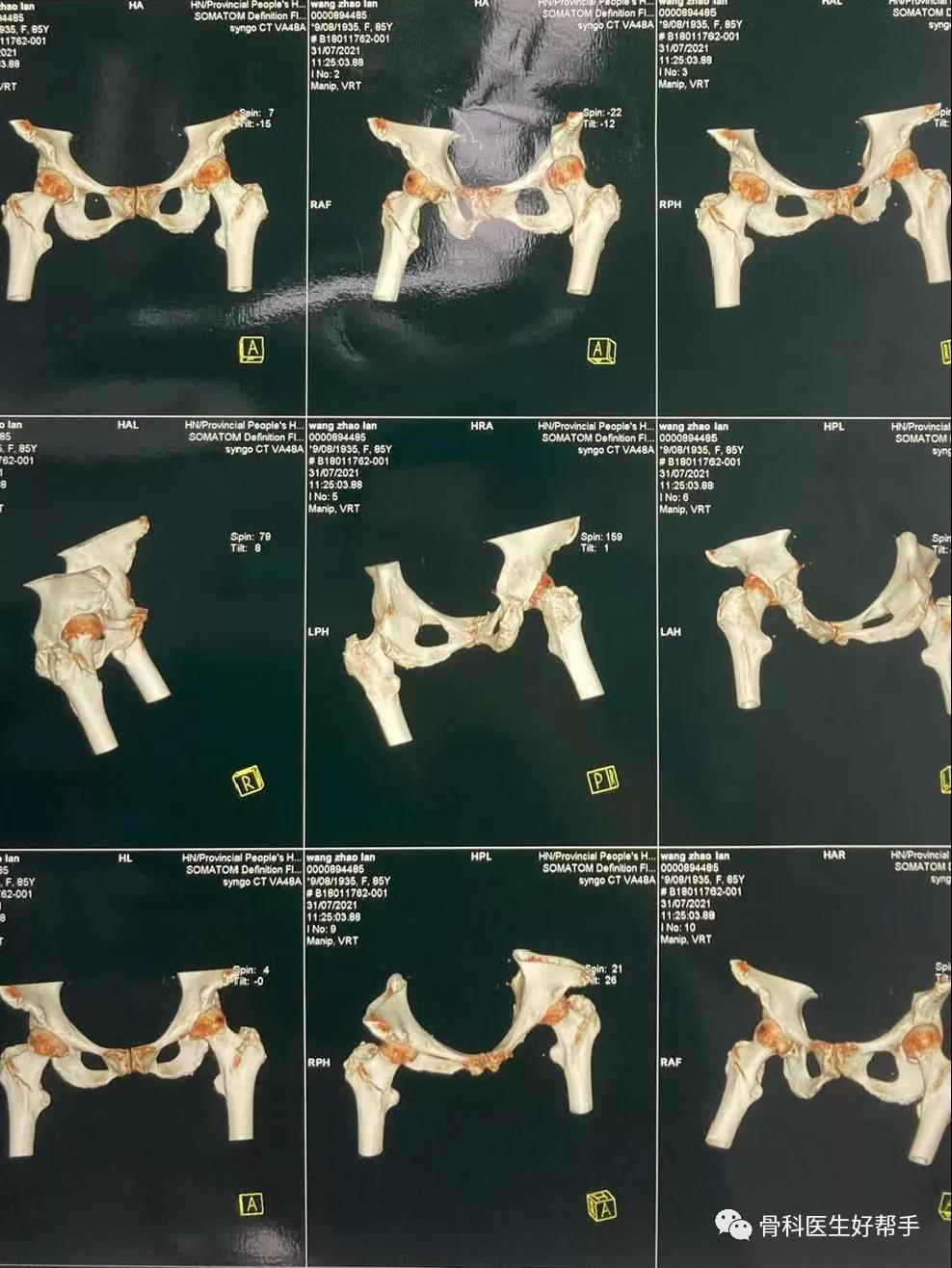
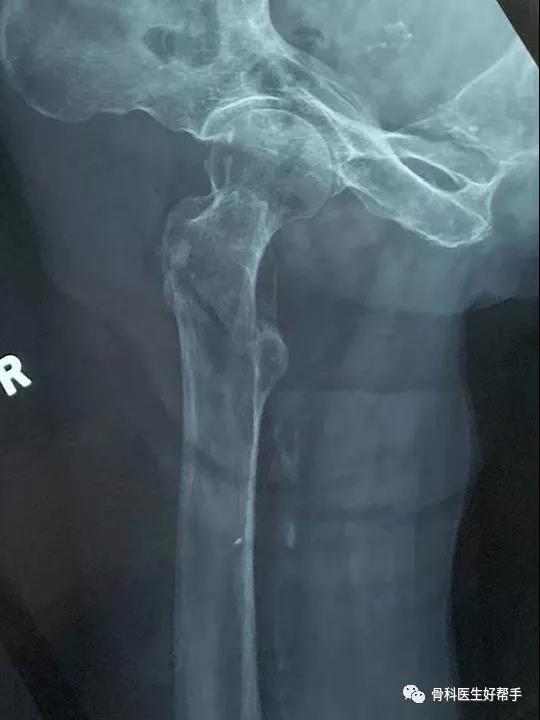
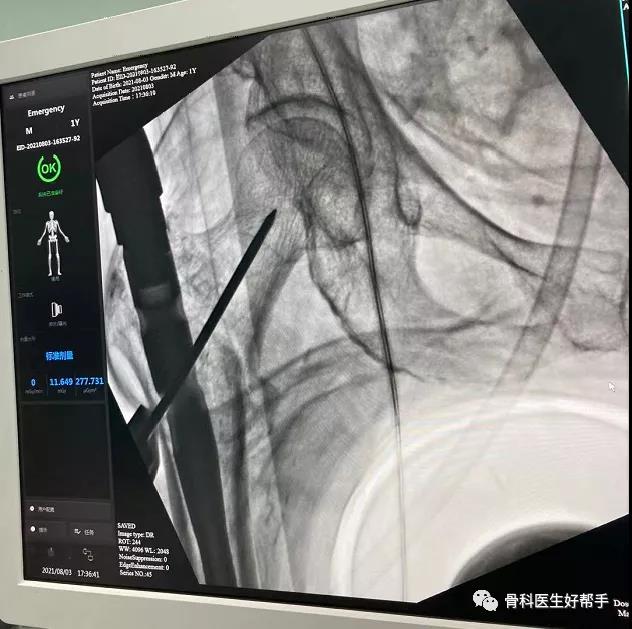
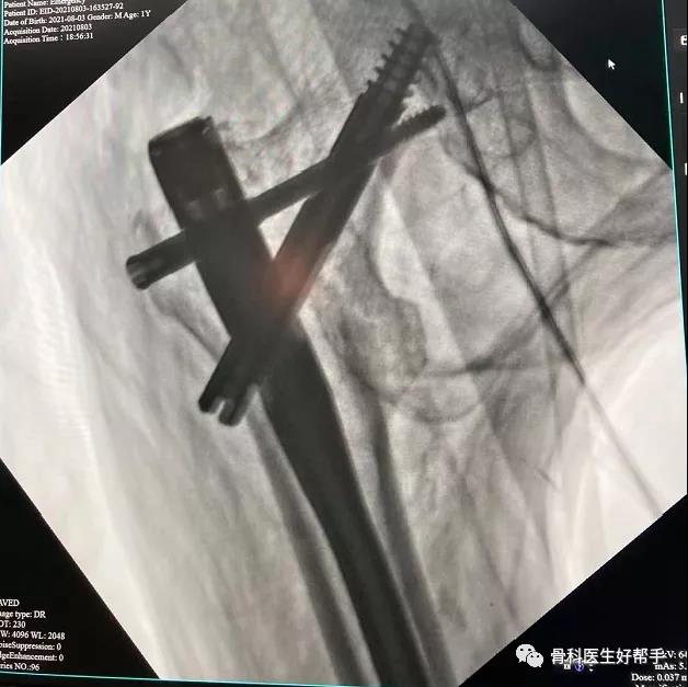
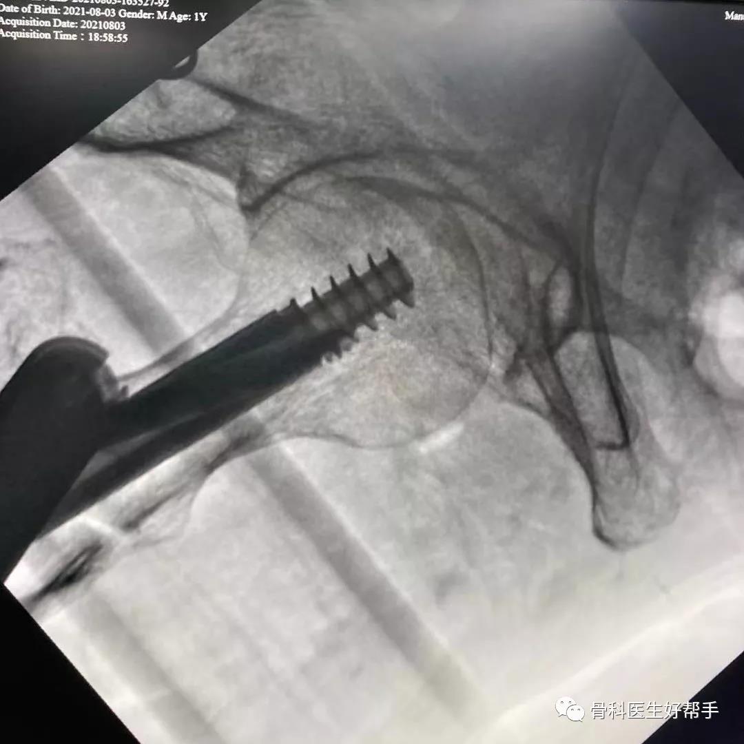
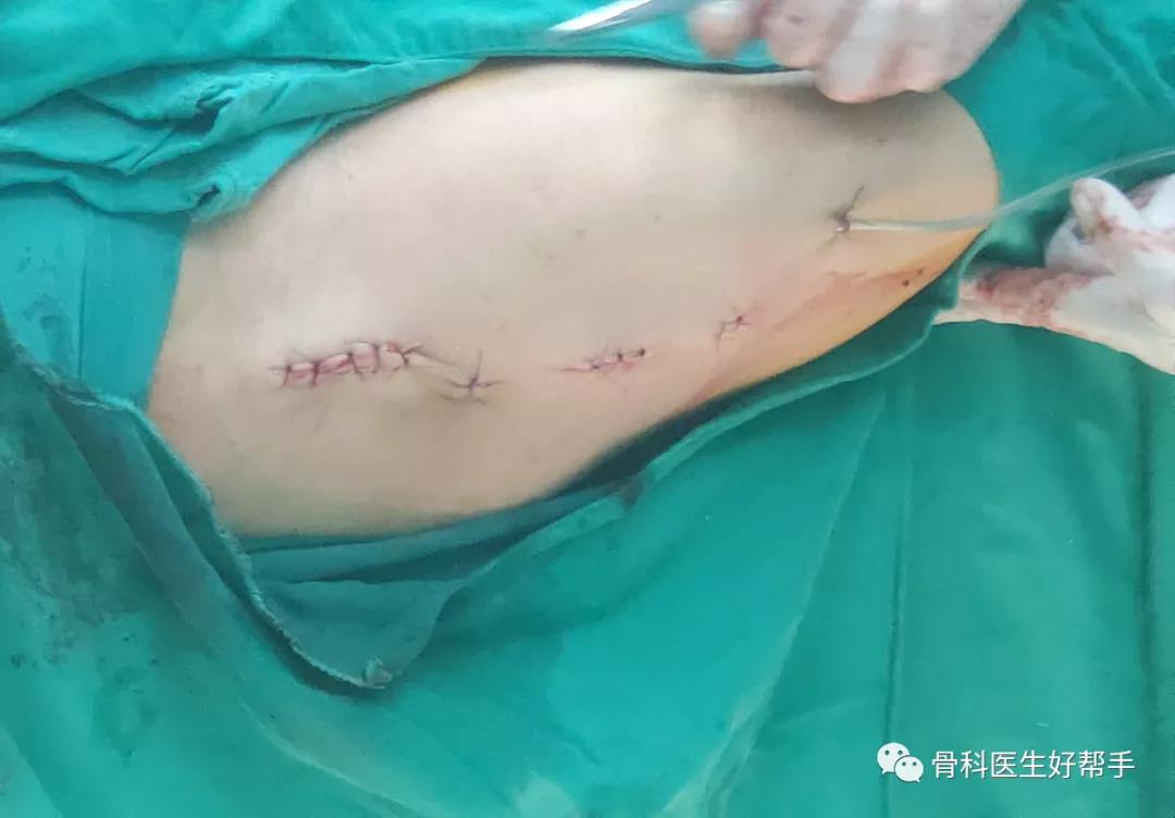
    Wang, Female, 85 years old, 1 hour before the visit, she was injured in an accidental fall, presenting with pain in her right hip joint and limited movement. An axial position of the right hip joint was performed in the emergency department, and the emergency treatment was admitted to the hospital as "right intertrochanteric fracture".

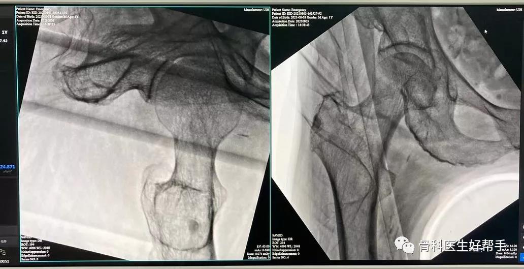
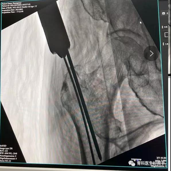
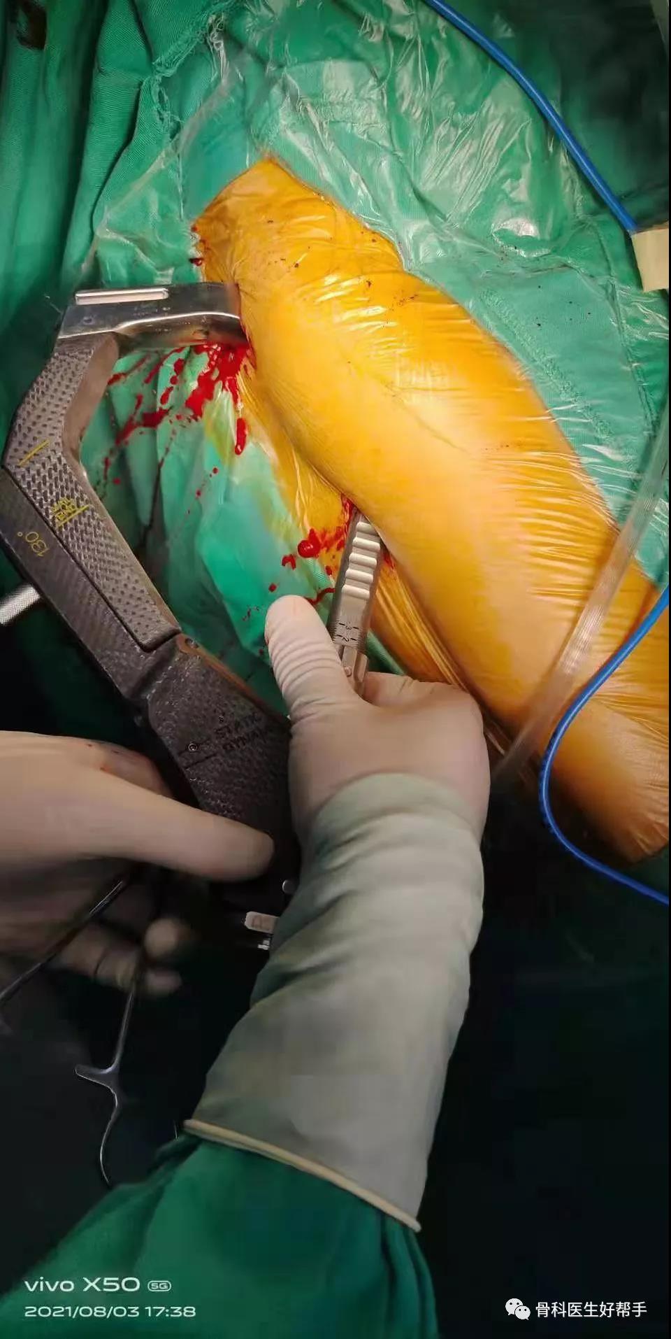
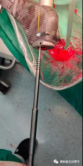
    Operation: closed reduction PFBN internal fixation

    Postoperative: fracture reduction, good effect of internal fixator.


    Checking Image:


    Intraoperative Images:


    Reduction:


    Guide wire position:


    Implantation:


    Postoperative Images:


  • Minimally Invasive Treatment of Achilles Fracture with Arthroscopic Technique Using Type II Heel Nail-in-Nail System

    Intra-articular calcaneal fractures are usually treated with traditional internal fixation involving a lateral widening incision followed by the use of calcaneal locking plates after resetting. While this method allows patients to return to work as soon as possible after the operation, it has several disadvantages. These include a large trauma caused by the L-shaped incision, the risk of skin necrosis, potential damage to the peroneal nerve during the operation, significant postoperative external adhesion, and limited mobility function. To avoid these complications, clinicians have started shifting towards minimally invasive internal fixation techniques.

    Minimally invasive internal fixation has proven to be effective in treating patients with intra-articular fractures of the heel bone. It involves smaller incisions, utilizes X-ray fluoroscopy-assisted resetting, facilitates surgical operations, minimizes soft tissue stripping to protect the surrounding blood supply, and provides a favorable biological environment for soft tissue repair and fracture healing. This approach significantly reduces the incidence of adverse reactions and shortens the operation time. In recent years, ankle arthroscopy has also been increasingly used for diagnosing and treating intra- or extra-articular diseases of the foot and ankle. The application of arthroscopic technology in heel bone fractures, assisted by arthroscopy, brings new hope and direction to treatment. we are orthopedic implants manufacturer,website: https://www.doublemedicalgp.com

  • Why Do Surgeons Use Intermittent Pneumatic Compression Devices During Surgeries


    Surgical procedures often involve prolonged immobility, which can increase the risk of developing common complications such as deep vein thrombosis (DVT) and pulmonary embolism (PE). To mitigate these risks, medical professionals frequently utilize intermittent pneumatic compression (IPC) devices. IPC devices have been proven to reduce and prevent blood clotting during and after surgery. In this article, we explore the reasons why IPC devices are an integral part of surgical care, their mechanism of action, and the benefits they offer.


    Reducing the Risk of DVT & PE
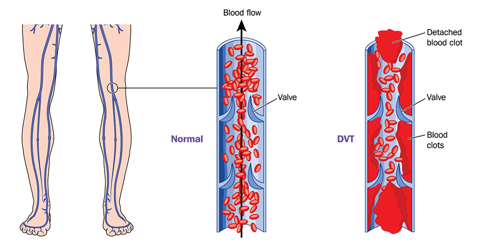


    DVT occurs when blood clots form in the deep veins of the legs, while PE involves the migration of these clots to the lungs. Both conditions can have severe consequences, leading to morbidity and mortality. During surgeries, patients may experience reduced blood flow and slower circulation due to sedation, immobility, and prolonged exposure to anesthesia. To counteract these effects, IPC devices are used to facilitate blood flow, preventing the formation of blood clots and reducing the likelihood of DVT and PE.


    How IPC Devices Work


    IPC devices comprise inflatable sleeves that are wrapped around the patient's legs or lower extremities. These sleeves are connected to an air pump, which periodically inflates and deflates the compartments, applying pressure on the veins. The alternating compression and release simulate the natural muscular contractions that occur during walking, thereby activating the calf muscle pump to propel blood back toward the heart.
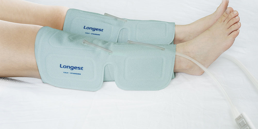


    Benefits of Using IPC Devices


    1. Prevention of Deep Vein Thrombosis: IPC devices significantly reduce the risk of developing DVT by increasing blood flow and preventing stasis in the lower extremities. By gently compressing the veins, these devices facilitate the removal of waste products and prevent the accumulation of clots, safeguarding patients from potentially life-threatening conditions.


    2. Enhanced Circulation: Prolonged periods of immobility can hamper blood circulation in surgical patients, leading to edema (swelling) and impaired tissue perfusion. IPC devices improve circulation by increasing venous return, thereby enhancing oxygen and nutrient delivery to tissues. This aids in the reduction of postoperative swelling and promotes faster healing.


    3. Decreased Hospital Stay and Costs: By using IPC devices, patients experience fewer postoperative complications such as DVT and PE. By mitigating these risks, the need for extended hospitalization and additional treatments can be reduced, resulting in shorter hospital stays and decreased healthcare costs.


    4. Nonpharmacological Intervention: IPC devices offer a non-invasive and nonpharmacological approach to preventing DVT and promoting blood circulation. This makes them suitable for patients with contraindications or allergies to certain medications, as well as those who prefer to minimize the use of drugs whenever possible.


    In conclusion, IPC devices are valuable tools in the prevention of postoperative complications, particularly DVT and PE. By enhancing blood flow, reducing stasis, and facilitating the calf muscle pump mechanism, IPC devices effectively reduce the risk of clot formation and improve circulation. When used during surgeries, these devices contribute to shorter hospital stays, reduced healthcare costs, enhanced patient outcomes, and improved overall patient safety. As medical advancements continue, coupled with the growing recognition of patient care, the use of IPC devices is expected to become even more widespread in surgical settings.


    Advanced IPC device for DVT Prevention
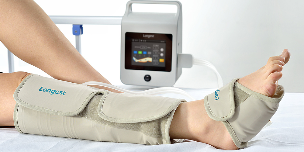


    The outstanding IPC device LGT-2200DVT is designed to make DVT prevention in hospital settings more convenient and more efficient. The device is remarkably compact in dimensions and lightweight, making the daily transport and usage of the device more convenient. Besides AC Power, the device can be powered by a rechargeable battery with over a 4-hour battery life, reducing the use of cords in the patient room to improve patient safety. 


    Despite its small size, it delivers impressive functionality with a high-performance pump and comprehensive compression sleeve options. A wide range of pressure allows us to meet more clinical needs and the comfort levels of different patients, improving clinical versatility and patient experience. 


    The LGT-2200DVT is a durable and reliable helper for healthcare givers in everyday DVT prevention work. It provides effective DVT prevention and management while ensuring patient safety and comfort.


    About Longest Medical


    Longest Medical has been committed to the development and manufacturing of advanced and reliable rehabilitation devices since 2000. The provide a wide range of rehabilitation products including IPC devices & sleeves for DVT prevention. Various models of DVT pumps that are user-friendly, lightweight, and portable are available. 

  • The Applications of Electrical Muscle Stimulation (EMS) Machine in Medical Aesthetics

    Electrical Muscle Stimulation (EMS) is a popular technology that has long been used in medical and rehabilitative settings for muscle strengthening, pain management, and physical therapy. In recent years, the EMS machine has gained significant recognition in the field of medical aesthetics for its potential to enhance cosmetic treatments and improve aesthetic outcomes. This article explores the various applications of EMS in medical aesthetics and examines how this technology is revolutionizing the industry.


    Non-Surgical Body Contouring

    EMS has emerged as a non-invasive technique for body contouring and muscle toning. This technology stimulates muscle contractions using electrical impulses, helping to improve muscle definition and promote fat reduction. It can be used to target specific areas of the body, such as the abdomen, buttocks, thighs, and arms, providing a non-surgical alternative to traditional liposuction and surgical body sculpting procedures.


    Facial Rejuvenation

    EMS is increasingly being utilized in facial rejuvenation treatments to improve muscle tone, enhance blood circulation, and promote collagen production. By stimulating the facial muscles, EMS helps to tighten and lift sagging skin, reduce the appearance of wrinkles and fine lines, and improve overall facial contours. This technology can be combined with other aesthetic treatments like dermal fillers and microdermabrasion to achieve optimal results.


    Postpartum Recovery

    EMS has shown promising results in postpartum recovery by helping women regain muscle strength and tone, especially in the abdominal area. After pregnancy, many women experience diastasis recti, a separation of abdominal muscles, which can be addressed with EMS. By stimulating the weakened muscles, EMS aids in repairing the separation and promoting the restoration of core strength.


    Cellulite Reduction

    EMS has been utilized as an effective method for reducing the appearance of cellulite. The gentle electrical impulses stimulate circulation, improve lymphatic drainage, and promote collagen synthesis. This helps to break down fat cells, smooth the skin's texture, and reduce the dimpled appearance associated with cellulite.


    Rehabilitation and Injury Recovery

    In addition to its aesthetic applications, EMS continues to be widely used in medical settings for rehabilitation and post-injury recovery. By targeting specific muscle groups, EMS can help strengthen weakened muscles, restore range of motion, and reduce pain. This therapy is often employed in cases such as muscle atrophy, joint injuries, and sports-related rehabilitation.


    The applications of Electrical Muscle Stimulation (EMS) machines in medical aesthetics are revolutionizing the field, offering non-surgical alternatives and enhancing the effectiveness of cosmetic treatments. From body contouring to facial rejuvenation and postpartum recovery, EMS has proven to be a versatile and effective technology. As further research and advancements continue, it is expected that EMS will play an increasingly significant role in medical aesthetics, providing patients with innovative solutions for their aesthetic concerns.


    Longest Medical EMS Solution


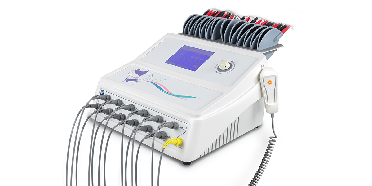
    LGT-2320BE is a 12-channel EMS machine designed for spa centers, weight loss centers, and beauty clinics to help their customers burn calories, tone muscles, and achieve their dream body faster and more safely.


    Features

    12-channels: treat more efficiently, improve productivity

    9 treatment modes: simplify operation steps and standardize treatment for consistency and comparison

    Reusable electrode pads: save costs and reduce wastes

    Electrode pad rack: better wire management

  • The Soaring Popularity of Shockwave Therapy in Aesthetic Treatments

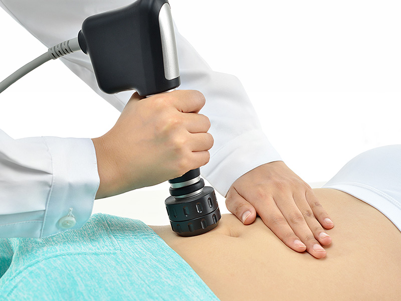
    Shockwave therapy for aesthetic treatment


    In recent years, shockwave therapy has emerged as a popular and effective treatment modality in the field of aesthetics. Originally used in medical settings for treating musculoskeletal conditions, shockwave therapy has now found its place in aesthetic clinics around the world. This article explores the skyrocketing popularity of shockwave therapy in aesthetic treatments, its mechanisms of action, commonly treated conditions, and the scientific evidence supporting its efficacy.


    WhatShockwave Therapy?

    Shockwave therapy, also known as acoustic wave therapy or extracorporeal shockwave therapy (ESWT), involves delivering high-energy acoustic waves to target tissues in a precise and controlled manner. These waves stimulate the body's natural healing response, improving tissue remodeling, collagen production, and blood circulation.


    Common Aesthetic Applications of Shockwave Therapy

    • Cellulite Reduction: Shockwave therapy has shown promising results in reducing the appearance of cellulite by targeting the fibrous connective tissue responsible for dimpling. The treatment helps to break down fibrous bands, enhance blood flow, and promote collagen production, resulting in smoother-looking skin.
    • Body Contouring and Fat Reduction: Combining shockwave therapy with other body contouring techniques, such as cryolipolysis or radiofrequency, has demonstrated enhanced outcomes in fat reduction and body shaping. The therapy aids in the breakdown of fat cells and improves lymphatic drainage, resulting in a more sculpted appearance.
    • Skin Tightening and Rejuvenation: Shockwave therapy stimulates collagen synthesis and remodeling, making it an effective option for skin tightening and rejuvenation. It can be used to address various cosmetic concerns, including fine lines, wrinkles, and sagging skin.


    Scientific Evidence and Studies

    Numerous scientific studies have investigated the efficacy and safety of shockwave therapy in aesthetic treatments:


    • A study published in the Journal of Cosmetic and Laser Therapy reported significant reductions in cellulite severity and improvements in skin texture and elasticity after shockwave therapy treatments.
    • Another clinical trial published in the Journal of Dermatological Treatment demonstrated favorable outcomes in body contouring and fat reduction when combining shockwave therapy with cryolipolysis, compared to cryolipolysis alone.
    • A review article in the Journal of Drugs in Dermatology summarized multiple studies and supported the use of shockwave therapy for skin tightening, cellulite reduction, and body contouring while emphasizing the importance of proper patient selection and treatment protocols.


    The Rise of Shockwave Therapy in Aesthetic Clinics

    The increasing popularity of shockwave therapy in aesthetic treatments can be attributed to several factors:


    • Non-Invasiveness: As a non-invasive treatment option, shockwave therapy offers minimal discomfort, no downtime, and a reduced risk of complications, making it appealing to individuals seeking non-surgical alternatives.
    • Versatility: Shockwave therapy can address multiple aesthetic concerns, making it a versatile tool in the aesthetic practitioner's toolkit. It can be used as a standalone treatment or in combination with other modalities, tailoring the approach to each patient's unique needs.
    • High Patient Satisfaction: Positive patient reviews and testimonials have contributed to the growing popularity of shockwave therapy. Many patients report visible improvements in their skin texture, cellulite appearance, and body contouring goals.


    Conclusion

    Shockwave therapy has gained widespread popularity in aesthetic treatments, offering non-invasive solutions for cellulite reduction, body contouring, and skin tightening. Supported by scientific evidence and positive patient feedback, this innovative therapy continues to attract both practitioners and individuals seeking effective, safe, and versatile aesthetic treatments. As technological advancements and research progress, the future holds great potential for further refining and expanding the applications of shockwave therapy in the field of aesthetics.


    Longest Medical - Your Reliable Shockwave Therapy Devices Supplier

    Longest Medical is a well-established medical and aesthetic devices manufacturer in China, with 23+ years of experience and expertise. Products are now exported to over 80 countries and regions.


    Our shockwave therapy devices are designed with simplicity, effectiveness, and comfort in mind, catering to different medical and aesthetic applications, including pain management, musculoskeletal disorders, cellulite reduction, etc. 

  • How to drape the four corners of the surgical drape?

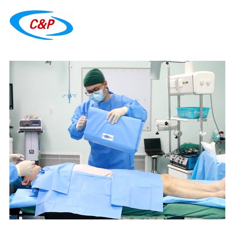
    The purpose of draping a surgical patient is to isolate the surgical site from other areas of the surgical site to help reduce the risk of surgical site infection. The order of draping the four corner surgical drapes sterile is as follows:

    1. Grasp the two corners of the drape and let it fall in front of you. Don't let it touch your body (unless you're wearing a sterile gown, which is fine) or anything else.

    2. Fold the top of the surgical drape over onto the side away from the body. This create a neat edge.

    3. Place your thumbs on the side of the drape closest to your body and wrap the drape around each hand. This prevents your hands from accidentally coming into contact with non-sterile areas of the patient.

    4. Always cover the side of the patient closest to your body first as this protects the gown from non-sterile tables.

    5. The surgical drape should move ("float") over the patient's body with the folded edges positioned at an appropriate distance from the intended incision line. Do not drag drapes over non-sterile areas of the patient's body. Once the drape is placed on the patient, only minimal adjustments are required. If the drape needs to be adjusted, move it only away from the sterile surgical field, never toward the sterile field.

    6. Repeat the process (steps 1-5) with the second drape - place it at right angles (90 degrees) to the first drape.

    7. Use a towel clip to secure the surgical drape to the patient. The towel clip should be placed at the intersection of the two surgical drapes. In living patients, remember that this is the first painful stimulus, so it is wise to inform the anesthetist that you are about to attach the clip.

    8. Tuck the towel clip under the upper drape. This will keep it out of the way (and prevent the suture material from getting caught in the clamps).

    9. Repeat the process with the third drape - place it at right angles to the first drape, on the opposite end of the sterile field from the second drape.

    10. Use towel clips to secure the drapes to the patient (this prevents the drapes from moving or shifting).

    11. When placing the final drape, either go to the other side of the table and place the drape, or sweep the drape over to the far side. Be careful not to contaminate the drapes through contact with accessories, team members, etc.

    12. After draping is completed, a surgical site of appropriate size will appear, surrounded by sterile surgical drapes secured with towel clips (all neatly tucked away).

    Notice:

    1. Remember the order in which you place the drapes: start from the near side (closest to your body), then place the drapes at both ends, and finally at the far side.

    2. If the surgical drape needs to be adjusted, you can only stay away from the sterile area and do not approach or cross the sterile area.

    3. When placing the clip, grab enough skin, but not too much - please pay attention to the patient.